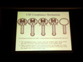
A CCSVI kezelése során a cél megszüntetni a véna szűkületet. Ezt általában egy felfújható ballonnal teszik, ami szétfeszíti a vénát. Kérdés, hogy a véna melyik pontján, mekkora átmérőjű ballonnal és mekkora nyomással.
Dr. Sinan (is) végzi a kuvaiti CCSVI műtéteket, valamint ő műt Egyiptomban is. Az ő technikája arra a felismerésre alapoz, hogy igazából az esetek nagy része visszavezethető a véna billentyűk problémáira, ezért magát a billentyű környékét tágítja első sorban. A billentyű körüli gyűrűszerű vénafal nem rugalmas, ezért annak tágításához nagy nyomásra van szükség. Sok esetben nem elegendő a tágítás, ha a billentyűk továbbra sem működnek (nyitnak) rendesen, akkor el kell roppantani ezt a gyűrűt, hogy kitágulhasson a véna.
Dr. Sinan részletes angol nyelvű angioplasztikai protokollját a cikk végén közlöm! (forrás)
Ezt a technikát alkalmazzák amerikában is már (San Diego, Dr. Arata), tekintettel dr. Sinan nagyon kicsi visszazáródási eredményeire.
Egy betegtársunk felvette a kapcsolatot dr. Sclafanival, és mint kiderült, őt is meggyőzte dr. Sinan, ő is ezt a technikát alkalmazza. Alant olvasható dr. Sclafani válasza a bennünk felmerült kérdésekre:
Kérdés: - Szeretném megtudni az On véleményet az alábbi kérdésekrol, melyek egy ideje elgondolkoztattak es egy kicsit meg is zavartak azzal az uj technikaval kapcsolatban amelyet Dr Sinan, es Dr Arata alkalmaz. ( Nagyobb meretu ballon, es a venabillentyuk szetroncsolasa )
Dr. Sclafani: - En ugyanezt a technikat alkalmazom. Dr Sinan volt az elso aki ezt az uj technikat leirta az altalam rendezett szimpoziumon meg tavaly júniusban. Meglepett, de egyben megelégedettsegel toltott el Dr Sinan kijelentese miszerint a betegek kepesek voltak toleralni a venak ilyen merteku tagitasat. Engem aggodalommal toltott el az ilyen nagy meratu ballonok alkalmazasa, melyet akkoriban problematikusnak gondoltam, mivel felteteleztem hogy trombizishoz vezet.
Kérdés: - Tudásom szerint a nyaki verőérben elhelyezkedo billentyu az egyetlen szelep a szív és az agy között , amely jelentos szerepet tolt be a venas reflux megelozeseben, valamint megakadalyozza a ver visszafolyásat az agy iranyaba. A beavatkozas soran valoban szetroncsoljak a venabillentyut, vagy pedig helyreallitjak annak eredeti, normal funkciojat?
Dr. Sclafani: - Megkerdojelezem ezeknek a venabillentyuknek a fontossagat. Az emberek 15%-ban ezek teljesen hianyoznak. A roncsolas attol fugg hogy milyen meretu a tagitas. A venabillentyu ket fo reszbol all: a korgyuru, mely az alapstrukturajat jelenti a szelepnek, valamint az ehhez kapcsolodo mozgatható vitorlak. A körgyűrűk rendkivul ellenalloak a tagitassal szemben, ami ertheto, hiszen ez tartja a vitorlakat a megfelelo tavolsagban egymastol( a verarammal szemben). Az ellenállásuk annyira eros, hogy 25 atmoszféra nyomás szukseges a szetfeszitesukhoz. Ezen folyamat soran ez a rugalmatlan szovet szétszakad.
Kérdés: - Abban az esetben, ha ezek a billentyuk vegervenyesen szetroncsolodnak ( es a ver szabadon aramlik), ez nem befolyasolja az koponyauri nyomast, ill. a reflux jelenseget?
Dr. Sclafani: - A megemelkedett központi venas nyomás atadodhat a jugularis venak teljes hosszaban olyan esetekben, ha nem kielegito a venabillentyuk mukodese, ill. amikor azok hianyoznak. Mint mar emlitettem a populacio 15 %-a nem rendelkezik ezzekel a billentyukkel, es ennek megsincs semmilyen kovetkezmenye. Mig megkerdojelezem ezeknek a venabillentyuknek a fontossagat az egyenesen jaro emlosoknel, addig viszont jol ertheto ezek szerepe a negy labon jaro allatoknal, ill. azoknal amelyek fejjel lefele toltik idejuk egy reszet, mint pl. a deneverek.
Mindezzel nem azt akartam mondani, hogy reflux nem fordulhat elo. Altalanossagban ez a nyomas szetszorodik a dural sinus teruleten es a nyakszirti kollateralis venakban. Nehany beszamolo alapjan viszont ezt az allapotot osszefuggesbe hozzak az atmeneti globalis amneziaval (TGA).
Mindezek ellenere ezt a potencialis lehetoseget nem szabad szem elol tevesztenunk. Amennyiben a jugularis vena kepes 16-18mm-es meretig kitagulni, de a benne levo korgyuru 6 mm-en fixalva van, akkor egyreszt lehetseges, hogy a jugularis vena valoban nagyon kicsi. A vena ilyenkor megdagad, hogy elbirja a belle levo veraramot, csakugy mint ahogy a gatak megtartjak a tavaszi aradaskor a megemelkedett folyo vizet. Ugyanakkor a folyon levo hodvar korlatozza a normalis atfolyast es ezzel a folyasirannyal ellentetes iranyu aradast idez elo.
Kérdés: - Lehet –e tudni, hogy mi a hosszu tavu hatasa annak, ha venabillentyuk vegervenyesen hasznavehetetlenne valnak? Emelkedik –e ezzel az atmeneti globalis amnezia kialakulasanak lehetosege?
Dr. Sclafani: - Igen lehetseges, de ezt altalaban a kozponti venas nyomas megemelkedesevel hozzak osszefuggesbe.
Kérdés: - Mi tortenik a venabillentyuk maradvanyaival?
Dr. Sclafani: - Velemenyem szerint tovabbra is a venafalhoz tapadtan maradnak. Mar lattam ilyet intravenas ultrahang keszulek segitsegevel, de egyenlore meg tobb tapasztalatra van szukseg az IVUS alkalmazasaban, mielott ezt behatobban tanulmanyozni tudnam.
Kérdés: - Abban az esetben, amikor a venabillentyuk a roncsolas utan mar nem leteznek lehetseges-e az, hogy az ez altal okozott atmeneti vervisszafolyas az agyba tovabbi SM leziok, ill. serulesek kialakulasahoz vezet?
Dr. Sclafani: - Addig amig korlatlan veratfolyas van a jugularis venakban, addig nem. De kerem ne felejtse el, hogy meg korai stadiumaban vagyunk a modszer kidolgozasanak
Itt pedig a részletes protokoll:
Step-by-Step Instructions for Angioplasty Treatment of CCSVI as Performed by Dr. Tariq Sinan’s Team
These notes were dictated to and written by Kathleen Lynch and were reviewed for accuracy by Doctor Tariq Sinan, Interventional Radiologist and Doctor Hussein Safar, Vascular Surgeon
Posted with the express permission of Dr. Tariq Sinan of Kuwait
http://www.drsinan.com/en/AboutMe.aspx
1. PRE-PROCEDURE MEDICATION:
Patient is given two 75 mg tablets of Plavix and a prophylactic dose of 1.5 grams of Zinacef intravenously 30 minutes prior to procedure.
2. SEDATION:
Foremost, the patient needs to be comfortable during the procedure, but it is best to use as little sedation as possible and concentrate on pain medication instead. The patient needs to be alert in case of neurological complications due to brain insult, and must be able to perform the valsalva maneuver, to respond to specific instructions regarding inspiration and expiration, and to answer the doctor’s questions. Over-sedation of the patient can interfere with their ability to do any of the above.
The patient’s apparent discomfort during ballooning can be informative to the doctor. A patient’s lack of discomfort usually indicates the need for a larger balloon. When pain medication is indicated, 25 mg of Fentanyl is administered just before inflation, resulting in a total of 75 mg. (25mg. for each jugular vein and 25mg. for the azygous vein). If the procedure is extensive, another 25 mg. may be administered at the discretion of the anesthetist.
3. ANTICOAGULATION:
Well-managed anticoagulation protocol is essential. For an adult male patient, a total of 5000 units of heparin is used, divided into three doses of 2000 units for each jugular and 1000 units for the azygous vein and administered intravenously. Dosage is adjusted to 4000 units intravenously for an adult female patient. A typical, complication-free balloon angioplasty of the jugular and azygous veins can be performed in approximately 120 minutes, but if complications or difficulties present and the procedure is extended an additional thirty to sixty minutes, another 1000 units of heparin is administered.
4. FEMORAL ACCESS:
Left femoral vein access is typically reserved for academic and investigative purposes, whereas right femoral vein access is indicated for treatment of jugular and azygous vein and valve abnormalities. A size 11 French guiding sheath is introduced and advanced, as it can accommodate most balloons and a wire at the same time. Start with a 4 or 5 French vertebral catheter with an angled Terumo wire with hydrophilic coating, 150 – 180cm in length. Sheath is 10 – 15 cm.
5. RIGHT INTERNAL JUGULAR VEIN:
Advance catheter to right internal jugular vein. The valves of IJV are just lateral and superior to the Acromio-clavicular joint. Navigate the valve of the RIJV by having patient perform the valsalva maneuver. Passage through the valve is easiest when it opens during expiration. At this point, do a “run” (contrast dye study) and assess for abnormalities during expiration and inspiration. It is crucial the abnormalities be viewed from an anteroposterior view. Sometimes an oblique view is needed. A regular J-tip 260cm wire is then introduced into the vein, and the catheter is withdrawn from the patient. Before dilation, a second wire is introduced using the vertebral catheter. Ideally, a J-tip stiff 260 cm wire should be used. There are now two wires in the RIJV; the regular Terumo wire and the stiff wire. Remove catheter and position balloon over the regular wire. Balloon size should be equal to vein size just cranial to the valve. The stiff wire remains outside the balloon. Advance the balloon to the valve and begin dilation. Balloon is to remain inflated for two minutes before deflating and dilating again. Repeat dilation for a total of five or six inflations, changing the position of the wire in relation to the balloon each time. The balloon will carry a “fingerprint” of the stenosis, and therefore should be repositioned so the “waist” of the balloon is in a different location in the vessel each time. The process should take at least twelve to fifteen minutes (five to six dilations at two minutes per dilation). Administer pain medication as needed. Any stenosis seen higher in the vein is not treated at this time. Check for complications with a contrast study. Withdraw from RIJV.
6. LEFT INTERNAL JUGULAR VEIN:
Advance catheter to LIJV and repeat the same procedure performed on the RIJV. Again, any stenosis seen higher in the vein is not treated at this time. Check for complications with a contrast study. Withdraw from RIJV.
7. AZYGOUS VEIN:
Using a left oblique view and a 100cm long C2 Cobra catheter and Terumo wire, advance to the azygous vein. The landmark for entry is the bifurcation of carina. If entry to the azygous vein is difficult, have the patient cough. Once in the azygous vein, perform a contrast study during inspiration and expiration to identify abnormalities. A single, regular 260 cm wire is introduced through the catheter into the azygous vein. A Cordis PowerFlex balloon is positioned onto the wire. Typically a 10cm x 4cm PowerFlex balloon is used for female patients, and a 12cm x 4cm PowerFlex balloon is used for male patients. Dilate each abnormality two or three times at two minutes per dilation. Continue until all abnormalities are addressed and treated. Upon completion, perform a contrast study during inspiration and expiration. Withdraw when satisfactory outcomes are achieved.
8. RETURN TO RIGHT INTERNAL JUGULAR VEIN:
Using the same vertebral catheter, return to the RIJV and perform a contrast study during inspiration and expiration to insure reflux is no longer in evidence. Upon evidence of even minimal reflux, repeat procedure (step five) with larger balloon. Withdraw from RIJV upon successful completion of dilation. Dilation is considered successful when there is no evidence of reflux.
9. RETURN TO LEFT INTERNAL JUGULAR VEIN:
Advance catheter to LIJV and perform a contrast study during inspiration and expiration to insure reflux is no longer in evidence. Upon evidence of even minimal reflux, repeat procedure (step five) with larger balloon. Withdraw from LIJV upon successful completion of dilation. Dilation is considered successful when there is no evidence of reflux.
10. POST-PROCEDURE:
Hand compression of incision site for 10 minutes.
Best rest.
Nothing per mouth for 4 hours.
Clexane injection four hours post-procedure (40 mg for females, 60 mg for males) Patient is taught self-injection
Patient discharged with medication and instructions. Plavix: 75mg once daily for 2 weeks; Clexane injections: 2x daily for 1 week; Aspirin: 75-80 mg. for 1 year.
Home rest for 24 hours.
No heat or stress exposure
Sleep in 45 degree inclined position (head higher than feet) for at least one month.
Hyperbaric Oxygen Therapy recommended.
Drink plenty of fluids.