 170 SM beteget vizsgáltak MRV-vel. Contrast-enhanced módszerrel 55%, time of flight módszerrel 61% -nál találtak áramlási problémákat. A kontroll csoportnál ezek a számok rendre: 5% és 10%. Forrás
Mégegy vadonatúj olasz kutatási anyag, a Zamboni protokollal 200 SM betegből 92%nál találtak CCSVIt. forrás ( 77 oldalon angolul összefoglalva)
Haacke doktor MRV protokollja elérhető, folyamatosan keres a kutatásaihoz csatlakozni vágyó - független és kíváncsi - orvosokat.
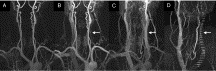
A comparative study of magnetic resonance venography techniques for the evaluation of the internal jugular veins in multiple sclerosis patients
- a Magnetic Resonance Innovations, Inc., Detroit, MI, USA
- b Pacific Interventionalists, Newport Beach, CA, USA
- c Wayne State University, Detroit, MI, USA
Abstract
Background and Purpose
The use of magnetic resonance imaging (MRI) to assess the vascular nature of diseases such as multiple sclerosis (MS) is a growing field of research. This work reports on the application of MR angiographic (MRA) and venographic (MRV) techniques in assessing the extracranial vasculature in MS patients.
Materials and Methods
A standardized MRI protocol containing 2D TOF-MRV and dynamic 3D contrast-enhanced (CE) MRAV was run for 170 MS patients and 40 healthy controls (HC). The cross-sectional area (CSA) of the internal jugular veins (IJVs) was measured at three neck levels in all subjects for both MRV techniques to determine the presence of venous stenoses. All data were analyzed retrospectively.
Results
For the values where both methods showed signal, the 3D method showed larger CSA measurement values compared to 2D methods in both IJVs, in both MS and HC subjects which was confirmed with student paired t-tests. Of the 170 MS patients, 93 (55%) in CE-MRAV and 103 (61%) in TOF-MRV showed stenosis in at least one IJV. The corresponding numbers for the 40 HC subjects were 2 (5%) and 4 (10%), respectively. Carotid ectasias with IJV stenosis were seen in 26 cases (15%) with 3D CE-MRAV and were not observable with 2D TOF-MRV. Carotid ectasias were not seen in the HC group. In the 2D TOF-MRV data, banding of the IJVs related to slow flow was seen in 58 (34%) MS cases and in no HC cases. MS patients showed lower average CSAs than the HC subjects.
Conclusion
The 3D CE MRAV depicted the vascular anatomy more completely than the 2D TOF-MRV. However, the 3D CE MRAV does not provide any information about the flow characteristics which are indirectly available in the 2D TOF-MRV in those cases where there is slow flow.
- CSA, cross-sectional area;
- HC, healthy control;
- IJV, internal jugular vein;
- CE, contrast-enhanced