HTML

CCSVI - a szklerózis multiplex vénás eredete

Egy olasz doktor, Paolo Zamboni felveti, hogy az SM (szklerózis multiplex) egyik fontos faktora lehet a nyaki vénás keringés elégtelensége. Eredményeit a világ számos országában alkalmazzák. Magyarországon még nem.
“Először átnéznek rajtad, aztán kinevetnek, aztán harcolnak veled, és végül nyersz.” (Mahatma Gandhi)
a blog szerzője elérhető: msccsvi@gmail.com

Linkblog

Friss topikok

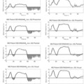
Új kutatási eredmények (2013 január)  alapjaiban változtatták meg az SM-ről alkotott képet. A legmodernebb MRI leletekkel bizonyított, hogy a szürkeállomány sérülése az SM legkorábbi szakaszában megtörténik és a megfigyelt vas kiválás, valamint atrópia (agyi oxigénhiányos állapot) mértéke összefügg a gyulladás mértékével:

... A number of clinical observations as well as recent neuropathologic and neuroimaging studies have clearly demonstrated extensive involvement of the thalamus, basal ganglia, and neocortex in patients with MS. Modern MRI techniques permit visualization of GM lesions and measurement of atrophy. These contemporary methods have fundamentally altered our understanding of the pathophysiologic nature of MS. Evidence confirms the contention that GM injury can be detected in the earliest phases of MS, and that iron deposition and atrophy of deep gray nuclei are closely related to the magnitude of inflammation. ... forrás: (pubmed link)

Hogy ez miért fontos? Erről már 2011 májusában közöltem egy posztot, lássuk újra:

Az SM diagnosztikája jelenleg főként MR készülékkel történik, a fehérállomány gócait térképezik fel. Ez azért van, mert a fehérállomány demielinizációját vagyunk jelenleg képesek megjeleníteni MR készülékkel. De talán nem látjuk a teljes képet...

Csak mostanában vagyunk képesek a szürkeállományt is vizsgálni MR-rel. De mi van ha eddig nem a megfelelő vizsgálatokat végeztük? Mi van ha az SM-nek nem a fehérállományban bekövetkezett mielinvesztés az oka, hanem a szürkeállományban bekövetkezett atrópia?

(A szürkeállomány az agy egy része, mely az agy oxigénszükségletének 94% -át használja el normál esetben. A teljes agy 40%-a a szürkeállomány és 60% a fehérállomány.)

Amikor az EAE modellt dr. Rivers létrehozta az 1930-as években,
http://www.ncbi.nlm.nih.gov/pmc/articles/PMC2212888/ akkor az emberi fehérállomány sérüléseit, gócait akarta egerekben modellezni: mivel csak azt ismerte az akkori tudásával. Ezt úgy érte el, hogy idegen antigéneket injekciózott az egerek agyába, ahol létrejött egy immun reakció, ami hasonlított az emberben - az emberi fehérállományban! - látottakra. Az összes mai SM gyógyszer úgy született, hogy az egerek EAE betegségét gyógyítják vele. És emiatt az emberekben nem is működnek olyan hatékonysággal… hogy miért? Mert az EAE és ADEM korántsem egyenlőek az emberi SM betegséggel… lássuk miért.

Az EAE jellemzően a fehérállomány betegsége, és nincsen benne nagyfokú szürkeállomány atrópia.

Az EAE egy valódi autoimmun betegség egerekben. Elfogadták, hogy ez az SM betegség egérmodellje. Ez a hibás feltevés oda vezetett, hogy az emberi SM betegséget is autoimmun betegségnek tartják, és ennek alapján készítik a gyógyszereket és végzik a kutatásokat. Ez a tévedés tragikus hatású.
http://www.mednat.org/vaccini/sclerosi.pdf

A progresszív SM betegségben nem figyelhetők meg fehérállomány gócok, nincsen mielin pusztulás, nincsen gyulladás… a betegség mégis romlik tovább. Miért? Hogy lehet hogy az SM "jól ismert" jelei nem igazak a progresszív formára, ennek ellenére a beteg állapota rohamosan romlik? Mi változik, amikor az SM progresszív lesz? Mi van ha SEMMI nem változik, és mi egészen eddig rossz tüneteket figyeltünk meg, mint például a fehérállomány gócok megjelenése? Hogyan lehet hogy egyes emberek 20 ilyen góccal az agyukban vígan bicikliznek, míg mások 2 góccal nem tudnak járni?

- "Eleinte az SM betegséget elsődlegesen fehérállomány központúnak gondoltuk - mondja Bianca Weinstock-Guttman neurológus - de mára már tudjuk, hogy a szürkeállomány sokkal inkább érintett mint a fehér." 
http://www.medicalnewstoday.com/articles/178458.php 

A következő tanulmányban a legmodernebb MR technológiával vizsgálták az SM betegek szürkeállományát. Arról szól, hogy a korai alacsony felbontású MR készülékekkel csak a fehérállományt lehetett vizsgálni, de ma már a modern készülékekkel láthatóak a "strukturális és funkcionális agyi károsodások" is.
A bizonyítékok sorra gyűlnek, hogy a kortikális demielinizáció és atrópia fontos faktor a progressziv SMben.
http://onlinelibrary.wiley.com/doi/10.1002/msj.20247/abstract

A következő tanulmányban a fehér és a szürkeállomány gócokat vizsgálták és arra jutottak, hogy szürkeállomány demielinizácója a fehérállomány demielinizációjától FÜGGETLENÜL zajlik le. Ez azért fontos, mert sok neurológus azt állítja, hogy először van a fehér, aminek következénye a szürkeállomány sérülés. Nem, ezek függetlenek:
http://archneur.ama-assn.org/cgi/reprint/64/1/76.pdf

A következő tanulmány arról szól, hogyan vizsgálható manapság a szürkeállomány sérülése SM-ben:

"A szürkeállomány gócok az SM legkorábbi fázisában is kimutathatóak és ezen gócok mennyisége és nagysága ÖSSZEFÜGG az SM beteg tüneteivel. (szerk. megj: nem úgy mint a fehérállománynál, ahol ezek nem függenek össze!) Ezek a szürkeállomány gócok egyben nagyon jó előrejelzői is az SM betegség jövőbeli várható állapotának.
http://www.mendeley.com/research/cortical-lesions-in-multiple-sclerosis/

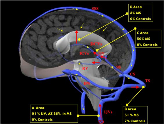
A következő képen az látható, hogy a Zamboni által talált vénás reflux hogyan érinti a szürkeállományt is:

Új eredmény: a következő videón a Hubbart Foundation által koordinált CCSVI tanulmányokból láthatjuk a legújabbak eredményeit. Dr. Giedrius Buracas ismerteti, hogy az általuk végzett fMRI BOLD vizsgálatok alapján a CCSVI érinti a kortikális területek oxigénellátottságát, sőt megmutatja, hogy a vénatágítás javít a szürkeállomány működésén!  konkrétan a 10. perctől látható ez:


A fenti prezentáció első része itt, a Hubbard Foundation összes videója itt érhető el.

Lassan fel kellene ismerni, hogy a közel 80 éves (!) egérmodell a továbbiakban már nem pontos, nem fedi le a jelenlegi tudásunkat az SMről. Tovább kellene lépni végre, fel kellene ébredni a 170 éves álomból és a tudományos tényeket kellene figyelembe venni, nem pedig ósdi egérmodelleket védelmezni. Az érvágás is kiment már a divatból itt az ideje, hogy az EAE is megtalálja a helyét: a nagy tévedések panoptikumában.

A gyógyszergyáraknak persze érdekük, hogy az EAE modellt védjék, hiszen erre tették a pénzüket, erre fejlesztik a gyógyszereket, és sok-sok év, évtized, amíg egy gyógyszer megtérül. De ez legyen az ő bajuk. Az internet segítségével immár hozzáférhetőek a legújabb kutatási eredmények és mi páciensek olvassuk ezeket és addig harcolunk amíg a mi érdekeink képviselete fontosabb lesz a profitnál.

A fordítás Joan Beal összzfoglaló cikke alapján készült.


Címkék: fmri tanulmány vizsgálat műtét hubbard ms ccsvi zamboni orvosoknak

5 komment

A bejegyzés trackback címe:

https://ccsvi.blog.hu/api/trackback/id/tr942905966

Kommentek:

A hozzászólások a vonatkozó jogszabályok  értelmében felhasználói tartalomnak minősülnek, értük a szolgáltatás technikai  üzemeltetője semmilyen felelősséget nem vállal, azokat nem ellenőrzi. Kifogás esetén forduljon a blog szerkesztőjéhez. Részletek a  Felhasználási feltételekben és az adatvédelmi tájékoztatóban.

The Ghost Wolf O1G · https://www.youtube.com/watch?v=Hy6Cf9nGNiw 2011.05.16. 07:51:56

Az Ashton Embry már régebben kifejtette egy írásában, hogy miért nem alkalmasak az EAE modellek az SM kutatására. Itt az anyag:

Experimental Allergic Encephalomyelitis:
A Misleading Model of Multiple Sclerosis
Subramaniam Sriram, MD,1 and Israel Steiner, MD2
Despite many years of intensive research, multiple sclerosis (MS) defies understanding and treatment remains suboptimal.
The prevailing hypothesis is that MS is immune mediated and that experimental allergic encephalomyelitis (EAE)
is a suitable model to elucidate pathogenesis and devise therapy. This review examines critically the validity that EAE is
an adequate and useful animal model of MS and finds credible evidence lacking. EAE represents more a model of acute
central nervous system inflammation than the counterpart of MS. We propose to reconsider the utilization of EAE,
especially when this model is used to define therapy. This will also force us to examine MS without the restraints
imposed by EAE, as to what it is, rather than what it looks like.
Ann Neurol 2005;58:939–945
Although the cause and pathogenesis of multiple sclerosis
(MS) are unknown, current prevailing hypothesis
favors MS to represent an autoimmune disorder directed
against nervous system antigens. 1–3 The basic
concept proposes that exposure to environmental
pathogens activates autoreactive T cells that recognize
central nervous system (CNS) autoantigens, leading to
inflammation and demyelination.4–7 This belief is promoted
by some similarities between MS and the various
animal models of experimental allergic encephalitis
(EAE).8
Since the initial experiments by Rivers, the stage was
set for the use of experimental animal models to study
CNS inflammation and demyelination.9 Over the last
30 years, the number of EAE-cited publications in English
has quadrupled; a Medline search identifies a total
of 678 articles on EAE between the years 1970 and
1980, 1,860 articles between 1990 and 2000, and approximately
1,600 publications since 2001.
Besides the utilization of EAE to study MS, it has
also been harnessed for developing therapeutic strategies
for MS.10–12 Indeed, the majority of the current
therapies being planned for phase II and III trials in
MS were first examined in EAE. Thus, EAE has become
a central player in the arena of MS. Is it indeed
a suitable and relevant research tool for MS? It has improved
our understanding of acute inflammatory demyelinating
syndromes, advanced our knowledge of
the genetic susceptibility to autoimmunity, and helped
uncover mechanisms of lymphocyte trafficking and the
role of blood–brain barrier in CNS inflammation. We
propose, however, that although EAE is a useful model
of acute human CNS demyelination such as acute disseminated
encephalomyelitis (ADEM), its contribution
to the understanding of MS has been limited. We focus
here on the lack of resemblance of the EAE model
with MS and examine its shortcomings when attempting
to extrapolate the findings from the model to the
human disease.
Experimental Allergic Encephalomyelitis, The
Prototypic Autoimmune Model
Myelin basic protein (MBP), proteolipoprotein (PLP),
myelin oligodendrocyte glycoprotein (MOG), myelinassociated
glycoprotein (MAG), and S-100 protein are
the major known CNS antigens that elicit an immune
response and cause paralytic disease in mice.13,14
MOG-induced EAE differs from MBP/PLP-induced
EAE in two major respects. Unlike MBP and PLPinduced
EAE, demyelination in EAE induced by
MOG is aggravated with concomitant administration
of anti–MOG antibodies, suggesting a prominent role
for a humoral response in the development of the inflammatory
pathology.15 Also, in some strains of mice,
the immune response to MOG is restricted by CD8
rather than CD4 T cells.16,17 Two additional myelin
antigens, MAG and CNP-ase are considered to be potential
autoantigens in MS because they can induce en-
From the 1Department of Neurology Vanderbilt Medical Center,
Nashville, TN; and 2Department of Neurology, Hadassah University
Hospital, Jerusalem, Israel.
Received Feb 15, 2005, and in revised form Jul 13 and Sep 28.
Accepted for publication Oct 8, 2005.
Published online Nov 28, 2005, in Wiley InterScience
(www.interscience.wiley.com). DOI: 10.1002/ana.20743
Address correspondence to Dr Sriram, Multiple Sclerosis Center,
1222H Vanderbilt Stallworth Rehabilitation Hospital, 2201 Children’s
Way, Nashville, TN 37212.
E-mail: subramaniam.sriram@ vanderbilt.edu
POINT OF VIEW
© 2005 American Neurological Association 939
Published by Wiley-Liss, Inc., through Wiley Subscription Services
cephalitis in animals. Studies of the presence of immune
response to MAG in MS patients are limited.18
Although the immune response to CNP-ase in MS patients
is as yet undetermined, cross-reactivity in response
to heat shock proteins and CNP-ase has suggested
a mechanism for induction of an autoimmune
response.19 S-100 is an astrocytic protein and immunization
of rodents produces an encephalitic picture with
only minimal demyelination.20
EAE is characteristically an acute monophasic illness
(as compared with the chronic relapsing course of MS)
making it more pertinent to ADEM. However, even
the development of chronic relapsing models of EAE
(CR-EAE) in rodents has not improved the relevance
of EAE in view of the continued differences between
the CR-EAE and MS.21–23
The Nature of the Inflammatory Response in
Experimental Allergic Encephalomyelitis and
Multiple Sclerosis
The ability of CD4 MBP-reactive T cells to induce
paralytic signs in mice established the immunological
basis of EAE. Immunohistochemical studies examining
the phenotype of the inflammatory cells have shown
the presence of T cells in both EAE and MS lesions.
24–27 However, CD4 T cells dominate the
perivascular regions of the inflammatory focus in EAE
induced by MBP and PLP. On the other hand, the
pathology of the demyelinating lesions in MS span a
spectrum between those that show prominent inflammation
and demyelination to others that represent an
oligodendrogliopathy with minimal inflammation of
demyelinated regions.28 In inflammatory MS lesions,
the predominant cells are macrophages and CD8 T
cells. CD4 T cells while present are infrequent.29–31
Isolation of T cells from MS brains by micromanipulation,
followed by targeted amplification of T-cell receptor
(TCR) genes showed a restricted expansion of
CD8 clones. Although CD4 clones were also isolated
from MS brains, they did not show a restricted TCR
expression pattern, suggesting they were not representative
of a clonally expanded population.32,33 In another
study, overrepresentation of CD8 T cells was
seen in spinal fluid of MS patients. These T cells, some
of the memory phenotype, were stable over several
months. In certain patients, the expansion of CD8 T
cells involved a restricted TCR V gene expression pattern
indicative of a clonal expansion.34,35 In the brain
parenchyma of MS patients, these CD8 T cells are
present in close apposition to the myelin membranes,
signifying that they may indeed play a role in tissue
damage.36 To further confound the issue, CD8-
restricted T-cell reactivity to MOG is sufficient to induce
EAE in mice, whereas there is little evidence of
CD8 T cells reactive to MOG peptides in MS.37,38
These findings question the relevance of the CD4 autoreactive
T-cell repertoire in the periphery of EAE lesions
to MS pathogenesis39 (Table 1).
Is Multiple Sclerosis a Th1-Mediated Disease as
Is Experimental Allergic Encephalomyelitis?
The separation of T-cell clones into two mutually exclusive
cytokine secretion patterns, Th1 and Th2,
evolved into dividing presumable inflammatory diseases
as being either Th1 (characterized by secretion of interleukin
[IL]–2, and -interferon) or Th2 (characterized
by secretion of IL-4, IL-5, and IL-13) mediated.40
Because Th1 cells are sufficient for adoptive transfer of
EAE, and -interferon is seen in MS lesions, it was
proposed that Th1 cells may be directly involved in
both MS and EAE. However, this is not true in all
EAE models and in every biological context: (1) MBPreactive
Th2 cell clones that secrete IL-4 and low levels
of -interferon also cause EAE41; (2) -interferon–deficient
mice developed EAE with greater severity after
immunization with either MBP or MOG peptides42;
(3) treatment of mice with -interferon caused attenuation
of disease and treatment of mice with anti–-
interferon antibodies induced worsening of EAE.43
A clinical study that reported worsening of the disease
in patients receiving -interferon has supported
the view that MS, like EAE, is a Th1-mediated autoimmune
disease. However, a careful reading of the report
raises several questions. Side effects such as fever,
myalgias, and arthralgias were noted in virtually all five
patients in the group of patients receiving high-dose
-interferon which subsided with discontinuation of
the drug.44,45 All of the exacerbations involved the
worsening of old symptoms, and at the completion of
the study there was no residual defect in any of the
seven patients with relapses. Whether corticosteroids
were given to the patients with relapses is unclear, and
the study was done at a time when magnetic resonance
imaging (MRI) scans were not readily available. Thus,
the transient neurological symptomatology induced by
-interferon could merely represent clinical decompensation
due to fever or the action of other cytokines
(“pseudorelapses”) and not necessarily the evolution of
a new inflammatory process. In other studies, induction
of -interferon has been observed after treatment
of MS patients with intravenous immune globulin
without any increase in the incidence of relapses. Also
administration of poly-ICLC in secondary progressive
MS, a known -interferon inducer, has not shown any
adverse effect in MS patients.46,47 Hence, assigning a
central role for Th1 cytokines in MS, which therefore
would serve as a major argument for the relevance of
EAE to MS, seems unfounded.
940 Annals of Neurology Vol 58 No 6 December 2005
Fundamental Differences in the Pathology
between Multiple Sclerosis and Experimental
Allergic Encephalomyelitis
In a manner analogous to that seen in EAE, the inflammatory
response in MS is thought to be mediated
by the trafficking to the CNS of autoreactive T cells
(see Table 1). Such a mechanism, sometimes referred
to as the “outside to inside hypothesis,” was recently
challenged by work by Barnett and Prineas48 and supported
by other studies.24,48–50 They noted the occurrence
of oligodendrocyte death as the very early and
perhaps the initial event in the pathology of the
plaque, even before development of inflammation.
These observations are by no means novel.51 Evidence
of early noninflammatory changes in the CNS has also
been suggested by imaging studies but not confirmed
histologically.52–57 However, normal-appearing white
matter on MRI may still contain microscopic evidence
of inflammation. This and other observations may interpret
the inflammation in MS as one of the following:
(1) an epi-phenomenon that follows areas where
the loss of myelin is large, such as in the vicinity of
large fiber tracts or (2) an attempt to fight a damaging
process that initiates oligodendrocyte death. Under
such a scenario, MS (unlike EAE) is not a disease that
is mediated by the entry of T cells from the periphery
but is caused by direct death and destruction of nervous
system structures including, to a large extent, the
destruction of myelin. Although the neurological consequences
of inflammation, induced by the destruction
of the oligodendrocyte-myelin unit cannot be ignored
and may contribute to morbidity, this concept will
foretell that long-term reduction in the inflammatory
response (with the use of either antiinflammatory or
immunosuppressive therapies) is unlikely to alter the
natural course of the disease.
The former view that MS is exclusively a white
matter disease was challenged and proved wrong by
histological and imaging studies. Indeed, axonal damage
and neuronal loss are common features of MS
and may be a direct consequence of inflammation or
because loss of trophic factors necessary to maintain
the integrity of the neural-axonal unit. Histological
and MRI studies have shown significant cortical and
axonal damage in MS that is not seen in EAE.58–60
Whatever the mechanism, demyelination in the cortical
gray matter mantle extends from the pial surface
to the gray white junction and spreads laterally over
several contiguous gyrii.58–60 Most importantly, these
areas of myelin loss lack an inflammatory response.
Similarly, large regions of the spinal cord around the
central canal showed loss of myelin with decrease of
neuronal structures.
Table 1. Immunopathology and Response to Therapy in EAE and MS
EAE MS
Pathology
Location of demyelination Predominantly, perivenous sleeves of myelin
loss in spinal cord and brain
Demyelination not restricted to perivenous
regions of white matter; extensive demyelination
of cerebral cortex in the absence
of inflammation is common
Location of lesions Dependent on the autoantigen used for induction:
inflammation dominates in lumbar
regions in MBP and PLP EAE and
brainstem in MOG EAE
Periventricular areas, cortical mantle,
brainstem, optic nerves, and upper cervical
cord; lesions are uncommon in
thoracic and lumbar regions
Phenotype of cellular infiltrate CD4 T cells (MBP and PLP EAE) activated
macrophages and few CD8 T cells
Activated macrophages and CD8 T cells
of a restricted clonotype
Cytokine predominance TH1 bias in MBP and PLP EAE; TH2 bias
worsens MOG EAE
Variable; no clear cytokine preponderance
CSF immunology Antibodies to myelin antigens present in CSF Antibodies to myelin antigens are infrequent
in CSF and do not constitute the
antigen specificity of oligoclonal bands
Effect of immunotherapies
 interferon Depends on route of administration and can
either worsen on ameliorate EAE
Worsening of inflammatory lesions unproven
 interferon Variable; can worsen EAE if given after immunization
Decreases relapse rate: effect on progression
modest
Anti–TNF antibody Reverses EAE Worsens MS
Anti–VLA-4 antibody Reverses EAE Decreases relapses; effect on progression
not known
Anti–CD4 antibodies Cures EAE No evidence of clinical efficacy on relapses
or progression
EAE  experimental allergic encephalomyelitis; MS  multiple sclerosis; MBP  myelin basic protein; PLP  proteolipoprotein; MOG 
myelin oligodendrocyte glycoprotein; CSF  cerebrospinal fluid.
Sriram and Steiner: Rethinking EAE and MS 941
Pitfalls in Extension of Immunotherapies from
Experimental Allergic Encephalomyelitis to
Multiple Sclerosis
The most disappointing aspect of EAE as a potential
model for MS is its almost total inability to point toward
a meaningful therapy or therapeutic approach for
MS (Table 2). The spectrum of agents and approaches
that showed promising results in EAE is immense and
range from turmeric (used in Asian cooking) and
Padma-28 (exotic natural drug found in health food
stores) to modern genetic manipulation of the immune
system with cytokines and antigen. Nevertheless, when
applied to the human “counterpart,” most, but not all,
of these therapies proved disappointing.61,62 Glatiramer
acetate represents the only drug currently in use
whose application in a clinical setting was first proved
useful in EAE.63 Glatiramer acetate is modestly effective
in reducing relapses but has not prevented the progression
of MS.64
The reasons for this failure are not only, as shown
here, that MS and EAE differ quite substantially, but
also that even from the larger, more comprehensive
picture, most of the evidence suggests that the EAE
models do not reflect the pathology of a progressive
disorder as MS. Moreover, the various EAE models are
dissimilar in their pathology and immunology to such
an extent that it is unclear why one EAE model will be
better served than another.
In clinical studies aimed at inducing antigen-specific
tolerance to a potential encephalitogenic autoantigen
such as MBP, either worsening of disease was noted or
there was no change in the clinical course.65,66 Induction
of oral tolerance in trials aimed to prove this point
were also a disappointment.67 Likewise, there was no
beneficial effect of anti-CD4 antibody therapy on the
progression of MS despite profound decrease of CD4
T cells in peripheral blood.68,69 Equally, examination
of the therapeutic approach of switching from a Th1 to
a Th2 profile in MS patients might prove a dangerous
experiment, because pathological studies of brains of
patients with MS show a Th2 response (presence of
antibodies and complement) in the most destructive of
lesions and glatiramer acetate induced a Th2 profile
only after prolonged in vitro cultures of lymphocytes.
31,70,71 All this puts into question the hope that
an immunosuppressive and/or antiinflammatory drug
are likely to have a significant impact on MS.72 The
reports of worsening of MS after bone marrow transplantation
may become disheartening proof.73,74
Conclusions
He who would distinguish the true from the false, must
have an adequate idea of what is true and false.—Baruch
Spinoza 1632–1677
The arguments we have presented should lead to
several conclusions: EAE is a disorder that differs im-
Table 2. Agents Successful in Treating EAE
Antibodies to T-cell surface antigens CD3,CD4,T-cellreceptor,CD2,IL-2R,IL-2R,CD24,CD40LCD28
Antibodies directed to antigen-presenting cells MHC class II antigens, CD40, B7-1 and B7-2, Fc receptor blockade
Antibodies to NK cells Anti–NK cell antibody, -Gal ceramide
Antibodies to adhesion molecules VLA-4, ICAM-1, LFA-1
Antibodies to cytokines IL-2, IL-6, IL-12, IL-15, TNF-, IL-1, IL-23
Antibodies to chemokines Anti–MIP-1— Rantes
Antiinflammatory cytokines IL-4, IL-10, TGF-, IFN-, IFN-, ?-IFN
Antagonists of signaling molecules Tyrphostins (inhibitors of JAK-Stat activation), lysofyline, inhibitors of
MAP kinase pathway, inhibitors of NF-B activation, Inhibitors of
iNOS activation, amsamycin, cholera toxin, AMPA antagonists, glutamate
receptor antagonists, IL-1 receptor antagonists
Activation of nuclear receptors PPAR- retinoic acid
Hormones Estrogen, progesterone, vitamin D, DHEA, leptin antagonists
Antibiotics Minocycline, rapamycin
Antimetabolites and immunosuppressants FK-506, cyclosporin, dyspergualin, corticosteroids, azathioprine, cyclophosphamide,
mycophenolate, bone marrow transplantation
Gene therapies Targeted delivery of IL-4, IL-10
Inhibitors of enzymes HOMG coreductase inhibitors (statins), COX-2 inhibitors
Peptides/proteins Oral myelin proteins, glatiramer acetate, myelin peptides (iv)
Food supplements Essential fatty acid, omega 3 fatty acid, curcumin, padma-28, fish oil
Small organic molecules Linomide, silica, sodium phenyl acetate, copper chelators (Nacetylcysteine
aminde), laquinamod, piperazylbutroxide, uric acid, dermatan
sulphate, amionoguanidine, cuprizone, roliprim, H-2 receptor
antagonists, indoleamine 2-3 deoxygenase, FTY-270, pentoxyfyline
Miscellaneous Incomplete Fruend’s adjuvant, BCG vaccination, Helminthic infections
AMPA  alpha-amino hydroxy methyl propionic acid; BCG  Bacille Calmette Guerin; DHEA  dehydro epi androsterone; EAE 
experimental allergic encephalomyelitis; HMG  hydroxymethyl glutaryl coreductase; IFN  interferon; IL  interleukin; iNOS  inducible
nitric oxide synthase; MAP  microtubule-associated protein; MHC  major histocompatibity complex; MIP  macrophage inflammatory
protein; TGF-  transforming growth factor-.
942 Annals of Neurology Vol 58 No 6 December 2005
munologically and pathologically between species, according,
in part, to the type of antigen used to induce
it and the species in which the model is tested. None
of the EAE models represent MS and they therefore are
imprecise methods to elucidate either the pathogenesis
or to develop therapeutic strategies in MS. In addition,
EAE is not a valuable vehicle to examine therapies: the
inability to apply the therapeutic successes of our findings
from the EAE model to the human condition is
one of the arguments against the autoimmune hypothesis
for the pathogenesis of MS.
We propose a much more careful use of EAE, especially
when this model is utilized to define therapy.
There are more than 100 compounds of proven efficacy
in EAE, and we believe that it is pointless to add
any more to this list (see Table 2). It may also be important
not to extrapolate successful therapies from
other dysimmune conditions in the hope that MS may
represent a variation on the theme of a common disease
mechanism.
We therefore are forced to examine MS without the
restraints of EAE, as to what it is, rather than what it
looks like. It would be interesting to ask the question
of how one could approach the disease if animal models
were unavailable, and the only recourse would be to
examine the clues offered by our patients and from relevant
genetic, imaging, and epidemiological studies in
humans. We believe that the current available pathologic
as well as radiological data would argue favorably
in examining issues outside of the “autoimmune hypothesis”
as central elements in the disease process.
We are grateful to Drs Nisipiano, Steiner-Birmanns, and Wirguin
for careful reading of the manuscript and useful suggestions and to
the families of J. Falker, P. Griffin, W. Weaver, T. West, and S.
Smith for their support of the MS Center.
References
1. Hemmer B, Archelos JJ, Hartung HP. New concepts in the
immunopathogenesis of multiple sclerosis. Nat Rev Neurosci
2002;3:291–301.
2. Noseworthy JH, Lucchinetti C, Rodriguez M, Weinshenker
BG. Multiple sclerosis. New Engl J Med 2000;343:938 –946.
3. Wekerke H. Immunology of MS. In: McAlpine’s multiple sclerosis.
3rd ed. New York: Churchill Livingstone, 1998:
379–407.
4. Zamvil S, Steinman LS. EAE and autoimmunity. Annu Rev
Immunol 1990;8:579–621.
5. Bright JJ, Sriram S. Immunotherapy of inflammatory demyelinating
diseases of the central nervous system. Immunol Res
2001;23:245–252.
6. Owens T, Sriram S. The immunology of multiple sclerosis and
its animal model, experimental allergic encephalomyelitis. Neurol
Clin 1995;13:51–73.
7. Sospedra M, Martin R. Immunology of multiple sclerosis.
Annu Rev Immunol 2005;23:683–747.
8. Raine CS. The Dale McFarlin memorial lecture. The immunology
of the MS lesion. Ann Neurol 1994;36:S61–S72.
9. Rivers TM, Schwentker FF. Encephalomyelitis accompanied by
myelin destruction experimentally produced in monkeys. J Exp
Med 1935;61:689 –701.
10. Steinman L. Assessment of animal models for MS and demyelinating
disease in the design of rational therapy. Neuron
1999;24:511–514.
11. Martin R, McFarland H. Experimental immunotherapies for
multiple sclerosis. Semin Immunopathol 1996;18:1–24.
12. Jyothi MD, Flavell RA, Geiger TL. Targeting autoantigenspecific
T cells and suppression of autoimmune encephalomyelitis
with receptor-modified T lymphocytes. Nat Biotechnol
2002;20:1215–1220.
13. Kies MW, Alvord EC. Allergic encephalitis. New York: C.C.
Thomas, 1959:293–299.
14. Pettinelli CB, McFarlin DE. Adoptive transfer of experimental
allergic encephalomyelitis in SJL/J mice after in vitro activation
of lymph node cells by myelin basic protein: requirement for
Lyt 1 2- T lymphocytes. J Immunol 1981;127:1420 –1423.
15. Genain CP, Abel K, Belmar N, et al. Late complications of
immune deviation therapy in a nonhuman primate. Science
1996;274:2054 –2057.
16. Huseby ES, Liggitt D, Brabb T, et al. A pathogenic role for
myelin-specific CD8() T cells in a model for multiple sclerosis.
J Exp Med 2001;194:669–676.
17. Sun D, Whitaker JN, Huang Z, et al. Myelin antigen-specific
CD8 T cells are encephalitogenic and produce severe disease
in C57BL/6 mice. J Immunol 2001;166:7579 –7587.
18. Schmidt S. Candidate autoantigens in MS. Mult Scler 1999;5:
145–160.
19. Rosener M, Muraro PA, Riethmuller A, et al. 2,3-cyclic nucleotide
3-phosphodiesterase: a novel candidate autoantigen in
demyelinating diseases. J Neuroimmunol 1997;75:28 –34.
20. Kojima K, Berger T, Lassmann H, et al. Experimental autoimmune
panencephalitis and uveoretinitis transferred to the Lewis
rat by T lymphocytes specific for the S100 beta molecule, a
calcium binding protein of astroglia. J Exp Med 1994;180:
817–829.
21. Gold R, Hartung HP, Toyka KV. Animal models for autoimmune
demyelinating disorders of the nervous system. Mol Med
2000;6:88 –91.
22. Rose LM, Richards TL, Petersen R, et al. Remitting-relapsing
EAE in nonhuman primates: a valid model of multiple sclerosis.
Clin Immunol Immunopathol 1991;59:1–15.
23. Lublin FD, Maurer PH, Berry RG, Tippett D. Delayed relapsing
experimental allergic encephalomyelitis in mice. J Immunol
1981;126:819–824.
24. Prineas JW. The neuropathology of acute MS lesion. Handbook
of clinical neurology 1985;3:213–257.
25. Raine CS. Multiple sclerosis: immunopathological mechanisms
in the progression and resolution of inflammatory demyelination.
Res Publ Assoc Res Nerv Ment Dis 1990;68:37–54.
26. Traugott U, Reinherz EL, Raine CS. Multiple sclerosis. Distribution
of T cells, T cell subsets and Ia-positive macrophages in
lesions of different ages. J Neuroimmunol 1983;4:201–221.
27. Traugott U, Reinherz EL, Raine CS. Multiple sclerosis: distribution
of T cell subsets within active chronic lesions. Science
1983;219:308 –310.
28. Lucchinetti C, Bruck W, Parisi J, et al. Heterogeneity of multiple
sclerosis lesions: implications for the pathogenesis of demyelination.
Ann Neurol 2000;47:707–717.
29. Booss J, Esiri MM, Tourtellotte WW, Mason DY. Immunohistological
analysis of T lymphocyte subsets in the central nervous
system in chronic progressive multiple sclerosis. J Neurol Sci
1983;62:219 –232.
Sriram and Steiner: Rethinking EAE and MS 943
30. Hauser SL, Bhan AK, Gilles F, et al. Immunohistochemical
analysis of the cellular infiltrate in multiple sclerosis lesions.
Ann Neurol 1986;19:578 –587.
31. Lassmann H, Ransohoff RM. The CD4-Th1 model for multiple
sclerosis: a crucial re-appraisal. Trends Immunol 2004;25:
132–137.
32. Skulina C, Schmidt S, Dornmair K, et al. Multiple sclerosis:
brain-infiltrating CD8 T cells persist as clonal expansions in
the cerebrospinal fluid and blood. Proc Natl Acad Sci USA
2004;101:2428 –2433.
33. Babbe H, Roers A, Waisman A, et al. Clonal expansions of
CD8() T cells dominate the T cell infiltrate in active multiple
sclerosis lesions as shown by micromanipulation and single cell
polymerase chain reaction. J Exp Med 2000;192:393– 404.
34. Cepok S, Jacobsen M, Schock S, et al. Patterns of cerebrospinal
fluid pathology correlate with disease progression in multiple
sclerosis. Brain 2001;124:2169 –2176.
35. Jacobsen M, Cepok S, Quak E, et al. Oligoclonal expansion of
memory CD8 T cells in cerebrospinal fluid from multiple
sclerosis patients. Brain 2002;125:538 –550.
36. Neumann H, Medana IM, Bauer J, Lassmann H. Cytotoxic T
lymphocytes in autoimmune and degenerative CNS diseases.
Trends Neurosci 2002;25:313–319.
37. Van der Aa A, Hellings N, Bernard CC, et al. Functional properties
of myelin oligodendrocyte glycoprotein-reactive T cells in
multiple sclerosis patients and controls. J Neuroimmunol 2003;
137:164 –176.
38. Koehler NK, Genain CP, Giesser B, Hauser SL. The human T
cell response to myelin oligodendrocyte glycoprotein: a multiple
sclerosis family-based study. J Immunol 2002;168:5920 –5927.
39. Ota K, Matsui M, Milford EL, et al. T cell recognition of an
immunodominant myelin basic protein epitope in multiple
sclerosis 1990;346:183–185.
40. Abbas AK, Murphy KM, Sher A. Functional diversity of helper
T lymphocytes. Nature 1996;383:787–793.
41. Lafaille JJ, Nagashima K, Katsuki M, Tonegawa S. High incidence
of spontaneous autoimmune encephalomyelitis in immunodeficient
anti-myelin basic protein T cell receptor transgenic
mice. Cell 1994;78:399–408.
42. Ferber IA, Brocke S, Taylor-Edwards C, et al. Mice with a disrupted
IFN-gamma gene are susceptible to the induction of experimental
autoimmune encephalomyelitis (EAE). J Immunol
1996;156:5–7.
43. Owens T, Wekerle H, Antel J. Genetic models of CNS inflammation.
Nat Med 2001;7:161–166.
44. Panitch HS, Hirsch RL, Haley AS, Johnson KP. Exacerbations
of multiple sclerosis in patients treated with gamma interferon.
Lancet 1987;1:893– 895.
45. Panitch HS, Hirsch RL, Schindler J, Johnson KP. Treatment of
multiple sclerosis with gamma interferon: exacerbations associated
with activation of the immune system. Neurology 1987;
37:1097–1102.
46. Bever CT Jr, Panitch HS, Levy HB, et al. Gamma-interferon
induction in patients with chronic progressive MS. Neurology
1991;41:1124 –1127.
47. Ling ZD, Yeoh E, Webb BT, et al. Intravenous immunoglobulin
induces interferon-gamma and interleukin-6 in vivo. J Clin
Immunol 1993;13:302–309.
48. Barnett MH, Prineas JW. Relapsing and remitting multiple
sclerosis: pathology of the newly forming lesion. Ann Neurol
2004;55:458–468.
49. Lumsden CE. The immunogenesis of the MS plaque. Brain Res
1971;28:365–371.
50. Trapp BD. Pathogenesis of multiple sclerosis: the eyes only see
what the mind is prepared to comprehend. Ann Neurol 2004;
55:455– 457.
51. Lumsden CE. Neuropathology of multiple sclerosis. In:
Koetsler JC, ed. Handbook of clinical neurology. Vol 3. New
York: Elsevier Science Publishing, 1972:217–309.
52. Matthews PM, Arnold DL. Magnetic resonance imaging of
multiple sclerosis: new insights linking pathology to clinical
evolution. Curr Opin Neurol 2001;14:279 –287.
53. Wolinsky JS, Narayana PA. Magnetic resonance spectroscopy in
multiple sclerosis: window into the diseased brain. Curr Opin
Neurol 2002;15:247–251.
54. De Stefano N, Guidi L, Stromillo ML, et al. Imaging neuronal
and axonal degeneration in multiple sclerosis. Neurol Sci 2003;
24(suppl 5):S283–S286.
55. Zivadinov R, Bakshi R. Role of MRI in multiple sclerosis I:
inflammation and lesions. Front Biosci 2004;9:665– 683.
56. Ranjeva JP, Pelletier J, Confort-Gouny S, et al. MRI/MRS of
corpus callosum in patients with clinically isolated syndrome
suggestive of multiple sclerosis. Mult Scler 2003;9:554 –565.
57. Fernando KT, McLean MA, Chard DT, et al. Elevated white
matter myo-inositol in clinically isolated syndromes suggestive
of multiple sclerosis. Brain 2004;127:1361–1369.
58. Bjartmar C, Wujek JR, Trapp BD. Axonal loss in the pathology
of MS: consequences for understanding the progressive phase of
the disease. J Neurol Sci 2003;206:165–171.
59. Bjartmar C, Trapp BD. Axonal degeneration and progressive
neurologic disability in multiple sclerosis. Neurotox Res 2003;
5:157–164.
60. Bo L, Vedeler CA, Nyland H, et al. Intracortical multiple sclerosis
lesions are not associated with increased lymphocyte infiltration.
Mult Scler 2003;9:323–331.
61. Badmaev V, Kozlowski PB, Schuller-Levis GB, Wisniewski
HM. The therapeutic effect of an herbal formula Badmaev 28
(padma 28) on experimental allergic encephalomyelitis (EAE)
in SJL/J mice. Phytother Res 1999;13:218 –221.
62. Natarajan C, Bright JJ. Curcumin inhibits experimental allergic
encephalomyelitis by blocking IL-12 signaling through Janus
kinase-STAT pathway in T lymphocytes. J Immunol 2002;168:
6506–6513.
63. Lisak RP, Zweiman B, Blanchard N, Rorke LB. Effect of treatment
with Copolymer 1 (Cop-1) on the in vivo and in vitro
manifestations of experimental allergic encephalomyelitis
(EAE). J Neurol Sci 1983;62:281–293.
64. Munari L, Lovati R, Boiko A. Therapy with glatiramer acetate
for multiple sclerosis. Cochrane Database Syst Rev 2004:
CD004678.
65. Bielekova B, Goodwin B, Richert N, et al. Encephalitogenic
potential of the myelin basic protein peptide (amino acids 83-
99) in multiple sclerosis: results of a phase II clinical trial with
an altered peptide ligand. Nat Med 2000;6:1167–1175.
66. Kappos L, Comi G, Panitch H, et al. Induction of a nonencephalitogenic
type 2 T helper-cell autoimmune response in
multiple sclerosis after administration of an altered peptide ligand
in a placebo-controlled, randomized phase II trial. The
Altered Peptide Ligand in Relapsing MS Study Group. Nat
Med 2000;6:1176 –1182.
67. Quinn S. Human trials: scientists, investors, and patients in the
quest for a cure. Perseus Publishing, Cambridge, MA: 2002.
68. van Oosten BW, Lai M, Hodgkinson S, et al. Treatment of
multiple sclerosis with the monoclonal anti-CD4 antibody cMT412:
results of a randomized, double-blind, placebocontrolled,
MR-monitored phase II trial. Neurology 1997;49:
351–357.
69. Lindsey JW, Hodgkinson S, Mehta R, et al. Phase 1 clinical
trial of chimeric monoclonal anti-CD4 antibody in multiple
sclerosis. Neurology 1994;44:413– 419.
944 Annals of Neurology Vol 58 No 6 December 2005
70. Ziemssen T, Kumpfel T, Klinkert WE, et al. Glatiramer
acetate-specific T-helper 1- and 2-type cell lines produce
BDNF: implications for multiple sclerosis therapy. Brainderived
neurotrophic factor. Brain 2002;125:2381–2391.
71. Dhib-Jalbut S, Chen M, Said A, et al. Glatiramer acetatereactive
peripheral blood mononuclear cells respond to multiple
myelin antigens with a Th2-biased phenotype. J Neuroimmunol
2003;140:163–171.
72. Weindl H, Hohlfeld R. Therapeutic approaches in treatment of
MS. Bio Drugs 2002;16:183–200.
73. Saiz A, Blanco Y, Carreras E, et al. Clinical and MRI outcome
after autologous hematopoietic stem cell transplantation in MS.
Neurology 2004;62:282–284.
74. Wiendl H, Hohlfeld R. Therapeutic approaches in multiple
sclerosis: lessons from failed and interrupted treatment trials.
BioDrugs 2002;16:183–200.
Sriram and Steiner: Rethinking EAE and MS 945

Sajnos az anyag csak egy pdf fileben van meg, de ha érdekel, szívesen elküldöm.

Ketel 2011.05.16. 09:01:59

Ezek a neurológusok hol vannak?Elnyomták őket? pl:Bianca Weinstock-Guttman neurológus ??Miért maradtak ezek az elméletek csak elméletek?Miért nem vizsgálták őket jobban meg?Apropó nekem is van atrophiám a homlok lebenyemben ezt a CT mutatta ki és az volt rá írva a tüneteket nem magyarázza ez az elváltozás.Ekkor jött az MR.Lényeg ami lényeg 74 referencia jelent meg ebben a témában/ha jól értelmezem/és semmi hatása?Miért siklanak el felette?Csak egy tárgy,kísérleti nyúl/nem egér/az ember??Tisztára Mendele jut eszembe róla,mivel különbek ezek az orvosok, gysz.cégek tőle???????Légyszi küld már el nekem az anyagot és megmutatom a neurológusomnak mit szól hozzá,úgy is megyek napokon belül hozzá.
Nagyon padlóra tett ez a cikk,még szúrni is felesleges magunkat?? Jó ezt eddig is gondoltam de most teljesedett be igazából...........

ccsvi · http://ccsvi.blog.hu 2011.05.16. 09:05:15

@Ketel: Senki nem mondta, hogy szúrni felesleges magunkat! Az SM gógyszerek működnek, a tüneteket enyhítik, DE nem az SM okát célozzák. Ezt a neurológusok is elismerik.

Ketel 2011.05.16. 10:42:44

@ccsvi: Egy betegségnek először az okát kellene megszüntetni!Mert ha csak a tüneteket kezeljük, nyomjuk el azzal semmit nem érünk.Jó azt tudom még senki nem tudja igazából az okát!S az is jó a betegeknek ha csökkennek a tünetek.DE a kór attól folyik tovább,rombol,ől, anélkül ,hogy tüneteket okozzon,vagy kisebb produktummal.Egyszer csak azt vesszük észre ,hogy nem is vagyunk!!Ezért kellene minden lehetséges elméleteket alaposabban megvizsgálni! Azt is tudom gysz-maffia az egész háttere.Lehet ,sőt többet ártunk a sm gysz-kel magunknak mint gondolnánk.Én is szurom magam ,csak ezen elgondolkodtam.Nekem is jó ha nincsenek,vagyis ha kevesebb a tüneteim,máris más szemmel nézem a világot.De sajnos nagyon ritka az olyan ,hogy jobb legyen a közérzetem.A neurológusok ezt elismerik a CCSVI ellen mégis fellépnek....na ez is a pénz...

Szilágyi Vilma 2015.02.24. 22:24:55

Kedves Balázs!
Angol tudásom nem "mives", de érdekel a folytatas!
Ha erdemben ertékes az írás, tudnal segíteni?
Köszönöm: Szilágyi Vilma
süti beállítások módosítása