HTML

CCSVI - a szklerózis multiplex vénás eredete

Egy olasz doktor, Paolo Zamboni felveti, hogy az SM (szklerózis multiplex) egyik fontos faktora lehet a nyaki vénás keringés elégtelensége. Eredményeit a világ számos országában alkalmazzák. Magyarországon még nem.
“Először átnéznek rajtad, aztán kinevetnek, aztán harcolnak veled, és végül nyersz.” (Mahatma Gandhi)
a blog szerzője elérhető: msccsvi@gmail.com

Linkblog

Friss topikok

a BREAKING NEWS -ok:

Az elmúlt napokban, hetekben nagyon fontos kutatási eredmények láttak napvilágot. Olyan kaliberűek, melyek alapján joggal lehet azt állítani: az SM kutatás alapjaiban változott meg 2015-ben.

1. Az emberi agyban felfedeztek limpatikus csatornákat. Igen, a nyájas olvasó - legyen az orvos vagy sokat olvasott beteg - most joggal kapja fel a fejét. 2015-ben derült ki, hogy az agyban vannak limpatikus csatornák, amikről eddig senki nem is tudott. Az atlaszokat újra lehet rajzolni. Bővebb infó és kapcsolat az SM betegséggel: http://ccsviinms.blogspot.hu/2015/06/rewrite-textbooks.html

2. Mai hír a Gladstone Institute -tól: Egyetlen csepp vér elegendő, hogy SMet okozzon. Mármint ha az agyba kerül. A Zamboni által sokat emlegetett vér-agy gát (gyakorlatilag egy vénafal) ha megsérül és vér jut az agyszövetek közé, akkor egy láncreakció során gyulladás és mielin pusztulást okozó immunreakció indul be. Ez megmagyaráz és bizonyít szinte mindent, amit Zamboni mondott a CCSVIről szerény véleményem szerint. Laikusoknak nincsen időm részletesen elmagyarázni ennek a hírnek a fontosságát. Kellően nyitott orvosoknak nem kell magyarázni. További infók erről a kutatási eredményről és kapcsolat az SMmel: http://ccsviinms.blogspot.co.uk/2015/10/a-single-drop-of-blood.html

a CCSVI blog VÉGE:

Azt hiszem ezekkel a hírekkel ennek a blognak az írását befejezem. Mintegy 7 éven keresztül követtem az SM kutatás legújabb híreit és igyekeztem a hazai orvostársadalom felé továbbítani. Nagyon sok rossz tapasztalatban volt részem, melyek nem vették el a lelkesedésemet. Hittem és hiszem a mai napig, hogy az SM és a keringési rendszer összefügg - ezt józan paraszti ésszel is egyértelműen látni lehet a fenti eredmények ismeretében.

Nincs értelme tovább írni ezt a blogot. Jómagam egyszerűen bizonyítottnak látom a Zamboni elméletet a Gladstone Institute fenti kutatási eredményével és ahogyan azt egy korábbi posztomban megígértem, gondoskodni fogok róla, hogy ennek a blognak a posztjai fennmaradjanak az örökkévalóságnak a magyar orvosok SM kutatásban betöltött szerepével együtt.

Természetesen azt is közölni fogom itt, ha végre elindul egy olyan magyar kutatási vonal, mely érdemben lesz képes végre foglalkozni a CCSVIvel.

Nekem most nincs több erőm, erőforrásom, időm ezt a blogot tovább írni. Remélem nem volt hiábavaló.

László, CCSVI beteg

msccsvi@gmail.com


From the Gladstone Institute Press Release:
In the current study, published in Nature Communications, the scientists created a new animal model of disease to determine if BBB leakage can cause autoimmunity. They discovered that injecting just one drop of blood into the brain set off the brain's immune response, kick-starting a chain reaction that resulted in inflammation and myelin damage.

These findings offer a completely new way of thinking about how the immune system attacks the brain--it puts the blood in the driver's seat of the onset and progression of disease," says senior author Katerina Akassoglou, PhD, a senior investigator at the Gladstone Institutes and professor of neurology at the University of California, San Francisco. "This opens up the possibility for new types of therapies that target blood coagulation factors, upstream of autoimmune processes."

"Our results provide the first evidence that blood promotes T cell responses against the brain," says first author Jae Kyu Ryu, PhD, a staff research scientist at the Gladstone Institutes. "Not only did we confirm that the presence of blood in the brain recruits peripheral immune cells to the area, which is sufficient to cause myelin destruction, we also identified fibrinogen as the critical protein driving this process."
"These findings question a long-held paradigm that myelin-specific T cells initiate inflammation in the brain through activation of microglia and brain macrophages," says Scott Zamvil, MD, PhD, a professor of neurology at the University of California, San Francisco and co-author on the paper. "This study demonstrates that the original paradigm may also occur in reverse. Namely, initial activation of microglia and brain macrophages may activate T cells."

1 komment

Három új SM gyógyszer is be lett jelentve az elmúlt hónapban. Mindhárom gyógyszer segített az SM betegeken stage I és stage II kutatásokban, lassította a progressziót, javította a tüneteket. Mindháromnak van egy új eposzi jelzője: neuroprotektiv. Ez most az új marketing jelző, amivel illetik ezeket a gyógyszereket, de ha kicsit jobban megnézzük milyen gyógyszerek ezek, akkor elég érdekes és ismerős dolgokat látunk.

A CCSVI teória szerint az SM nem más mint egy krónikus oxigényhiányos állapot melynek során az agy vérellátása lassul, kevesebb oxigént kap, lassul a véráram. Ennek oka a vénák szűkülete, elszűkülése, melynek  következtében olyan reakciók zajlanak le az agyban mint stroke betegek esetében, erről írtunk már többször is.

Ez a három új gyógyszer hasonló módon hat. Mindhármat évek óta használják stroke betegek és érrendszeri problémák esetében. Mindhárom az endotéliumra hat (ez gyakorlatilag a vénák fala) segít a vénafal ellazításában (nitric oxide), növelésében, csökkenti a vérnyomást.

Mindhárom tehát tágítja a vénákat, növeli a keringő vérmennyiséget az agyban.

guanabenz-- ellazítja az ereket, a vér könnyebben áramlik.
http://www.mayoclinic.org/drugs-supplements/guanabenz-oral-route/description/drg-20064106

ibudilast-- növeli az agyban a keringő vér mennyiségét, tágítja az ereket
http://www.ncbi.nlm.nih.gov/pubmed/18677969

biotin---csökkenti a vérnyomást, növeli a véráramlás mennyiségét, a stroke utáni alacsony oxigénszint kezelésére használják
http://www.ncbi.nlm.nih.gov/pubmed/18179728
http://www.google.com/patents/WO2014016003A1?cl=en

Mintha a vénatágítást gyógyszerrel végeznék el. A (valódi) SM kutatók tehát megértették Zamboni elméletét, mely szerint a véráramlással van gond az SM betegeknél. A gyógyszergyárak pedig pénzt fektettek ezekbe a kutatásokba, tehát nagyon alapos indokuk lehet.

Lehetne még hosszasan gondolkodni azon, hogy miért nem hagyják, hogy a betegek kitágítsák a vénájukat, vagy miért nem az egészséges életvitelt, mozgást, oxigénterápiát stb. javasolják, hanem helyette miért drága gyógyszereket dobnak piacra.

Várható volt, hogy a gyógyszeripar gyorsan fog kapcsolni, és elkezdenek Zamboni elmélete köré valamiféle gyógyszergyárat építeni. Reméljük legalább hatásos lesz valamennyire. Már az sem nagyon zavar ,ha hülyének néznek és a betegségemből élnek, csak segítsenek rajtunk végre.


Szólj hozzá!

isnvd5thlogo-c.pngVéget ért az ISNVD 2015-ös konferencia, ahol bemutatták a legújabb tudományos eredményeket, melyek a véráramlás és az idegrendszer kapcsolatáról szólnak. Mivel ez főként orvosokat érdekel és az anyag szerencsére hatalmas, ezért nem tudom még csak összegezni sem, vagy az összegzéseket lefordítani sem.

Itt letölthetők a 2015-ös absztraktok (pdf).  Ami jól látható számomra: a CCSVI kutatás gőzerővel zajlik és új területek nyílnak. Íme egy rövid összegzés Joan tollából:

2D and 3D analysis of vessels in the retina and the brain
Prof. Bart ter Haar Romeny, Ph.D.1,2
1Eindhoven University of Technology, Eindhoven, the Netherlands 2Northeastern University, Shenyang, China
This research is using the imaging of blood vessels in the eye's retina to get a picture of how blood is circulating throught the central nervous system. The blood vessels in the retina give an early picture of how brain diseases and breakdown of the blood brain barrier might be developing. Scanning the eye is easier and more cost-effective, as well.

Venous dysfunction and neurodegenerative diseases
Chih-Ping Chung MD PhD
Taipei Veterans General Hospital, National Yang Ming University, Taipei, Taiwan
Dr. Chung and his group have been studying the venous vasculature in relationship to brain disorders for over a decade. He has spoken at a few of the ISNVD conferences now. His new research focuses on how venous abnormalities are linked to white matter changes and how venous drainage impairment leads to dysfunction in Alzheimer's Disease.

 
Blood storage within the intracranial space and its impact on cerebrospinal fluid dynamics

Clive B Beggs 1, Simon J Shepherd 1, Pietro Cecconi 2 and Maria Marcella Lagana 2Medical Biophysics Laboratory, University of Bradford, Bradford, BD7 1DP, UK  Fondazione Don Carlo Gnocchi ONLUS, IRCCS S. Maria Nascente. Milan, Italy 
This study measured blood flow throughout the cardiac cycle by using MRI to visualize the flow in the necks of 14 healthy adults.  This study found that it is cerebrospinal fluid (CSF) which controls the volume changes inside the brain.  CSF interacts with the cortical veins to facilitate how much blood is stored.  This is an important finding, because any disturbance in venous outflow of blood and CSF will change the blood storage inside the skull, potentially leading to reduced cerebral circulation.

Advances in Treatment Strategies of Extracranial Venous Disease
Hector Ferral, MD
Senior Clinical Educator NorthShore University Health System
Dr. Ferral has been treating people with CCSVI for a few years now. He has also been an attendee and presenter at the ISNVD before. His new presentation will be looking at the technical advances being made in vein measurement and treatment of CCSVI.

Imaging of Brain Microvascular Disorders: lessons from the CADASIL model.
Hugues Chabriat, MD PhD;Department of Neurology, GH Lariboisiere, APHP, INSERM UMRS1161, University Paris 7 Denis Diderot, Paris France.
This research uses imaging to look at how the small vessel disease related to stroke and dementia develops and progresses.

Endothelin- 1 as a potential target for chronic brain hypoperfusion
Jacques De Keyser, MD, PhD, Free University of Brussels (VUB), Department of Neurology, Brussels, Belgium
This study is near and dear to my heart, as it is looking at how ET-1, a marker of endothelial dysfunction, is related to slowed blood flow in the brain, called hypoperfusion. People with MS have much higher levels of ET 1 in their blood than normals, and much slower cerebral blood flow. We also see this marker elevated in a number of neurodegenerative diseases, like Alzheimer's. In this study, the researchers used bosentan, a blood pressure medication, to treat ET 1 levels. This treatment increased cerebral blood flow in pwMS, and lowered ET-1 levels. (But bosentan has side effects, and is not easy on the liver. Much better, in my opinion, to lower ET-1 levels by addressing endothelial dysfunction, through diet, exercise, and lifestyle.)

TBI and hemodynamic changes in the brain

James R. Stone, MD, PhD
This presentation is looking at how traumatic brain injury induces ischemia, or a low-oxygen state, in the brain. TBI also changes cerebral blood flow and can cause a break in the blood brain barrier, igniting the immune system. New research is showing how explosive devices can cause TBI, even without direct physical contact.

Ultrasound contrast imaging of brain hemodynamic and perfusion Marcello Mancini, M.D.
Institute of Biostructure and Bioimage – CNR
Naples, Italy
This presentation will be looking at how new MRI and ultrasound technologies are allowing researchers to view cerebral circulation in MS.  People with MS show signs of hypoxia (low oxygen) injury and thrombosis (small clots) in the small veins of the brain. New technologies are allowing us to see that cerebral transit time is slowed in MS. 
 
Imaging of the Microvasculature
E. Mark Haacke, PhD 
Dr. Haacke, a presenter at all of the ISNVD conferences, returns this year to discuss how his invention of susceptibility weighted imaging (SWI) and MRA can be used to study the neurovascular system, and clarify the relationship between the venous sytem and CSF.


Update in computational fluid modelling of the brain
Mauro Ursino
This presentation is using mathmatical and computer models to simulate the complex mechanisms affecting cerbral circulation. Using these models shows how postural changes and stenosis in extra cranial arteries and veins can change upstream intercranial circulation.

Clinical Applications of Venous Treatment
Dr. Michael Dake, Stanford University
Dr. Dake, last year's ISNVD president and a founding member, will be presenting on the contributions of 2014 studies which have enhanced understanding of how endovascular and open surgical treatment of venous abnormalities has affected patients with MS, Alzheimer's disease, Parkinson's, POTS and other pathologies.
 
The Heart Brain Connection
MJ Daemen
Dr. Daemen is the keynote guest speaker. He is a neurocardiologist, a member of a new field of experts who are bringing together an understanding of how the heart and brain affect each other. As a member of the Dutch Heart Foundation, his group is looking at how cardiovascular disease is influencing cognitive function and cerebral circulation.

A NOVEL SONOGRAPHIC METHOD FOR REPRODUCIBLE JUGULAR VEIN PULSE WAVE ASSESSMENT
Paolo Zamboni, Francesco Sisini, Erica Menegatti, Giacomo Gadda, Mirko Tessari, Mauro Gambaccini
Vascular Diseases Center, University of Ferrara, Ferrara, Italy 
Dr. Zamboni's group is using ultrasound in the B mode (brightness mode) to measure the pulse wave in the jugular vein.  The way that the wave form looks is currently used to monitor for heart disease, however Dr. Zamboni's group is using this technique to find CCSVI.

Is there a role for mast cells dependent synthesis of Endothelin-1 in neurodegenerative diseases?
Pedro D’Orléans-Juste-1, Louisane Desbiens, Denis Gris-2
Departments of Pharmacoly-1 and of Pediatrics-2, Faculty of Medicine, Université de Sherbrooke, Sherbrooke, PQ, Canada
We know that levels of ET-1 (a marker of endothelial dysfunction) and mast cells (tissue cells of the immune system) occur in higher levels in people with MS.  This study uses the mouse model of EAE as well as a human isoform to study how mast cells found in the vicinity of spinal lesions are involved in the synthesis of ET-1.
 
Venous abnormalities in Meniere's Disease
P.M.Bavera; P. Cecconi; D. Alpini; F. Di Berardino 
This presentation will be using slides to show the correlation and differences between Meniere's Disease and MS, in regards to CCSVI imaging.  There are specific characteristics to the venous abnormalities seen in Meniere's.
 
Advances in Idiopathic Intracranial Hypertension Pathogensis: a Focus on Sinus Venous Stenosis
Roberto De Simone, Angelo Ranieri
Headache Centre  Dpt. of Neurosciences, Reproductive Sciences and Odontostomatology University of Naples “Federico II”
This research is focusing on how stenosis of the venous sinus is related to idiopathic intracranial hypertension (IIH)  There is a feedback loop which appears to occur in this situation.  The venous sinus collapses, cerebrospinal fluid pressure builds, which creates more compression.  Endovascular stenting of the venous sinus is a currently approved treatment to end this cycle of stenosis and hypertension.

In Endothelial function, the glymphatic system and New Drug Development
Endothelial dysfunction in neurodegenerative disease

J. Winny Yun, Emily Stevenson, Seiichi Omura, Fumitaka Sato, Ikuo Tsunoda, Alireza Minagar, Felix Becker, Trevor Castor, Adam Xiao, J. Steven Alexander, LSUHSC-Shreveport Molecular and Cellular Physiology, Microbiology, Virology, Neurology, Shreveport, Louisiana, USA.
Dr. Steven Alexander from LSU returns to the ISNVD to again discuss the endothelium in neurovascular disease. There is a vascular association of specific biomarkers found in MS. This new research is looking for a means to regulate these neurolymphatic markers, to help those with neurovascular diseases.

Fluid Dynamic Influences on Cerebrovascular Endothelial Activation Responses
Dr. Alexander and the LSU team look at how blood flow over endothelial cells affect their health. Laminar shear stress (regular blood flow) over endothelial cells is essential to their function. Disrupted flow causes endothelial cells dysfunction and death. Shear stress alterations could lead to a break down of the endothelial layer in the brain, and create a disturbance in the blood brain barrier and inflammation.

Cardiovascular risk factors and neurodegenerative disorders
Dr. Robert Zivadinov
Buffalo Neuroimaging Analysis Center, Department of Neurology, University at Buffalo, State
This research finds an association with cardiovascular risk factors (smoking, obesity, inactivity, high blood pressure) and MS.  MS patients who had one or more CV risk factors had higher lesions loads and more brain atrophy.


-------------------------------------------------------------------------


A 2014-es kutatási anyagok:

banner2.png

Véget ért az ISNVD 2014 -es konferencia, ahol bemutatták a legújabb tudományos eredményeket, melyek a véráramlás és az idegrendszer kapcsolatáról szólnak. Mivel ez főként orvosokat érdekel és az anyag szerencsére hatalmas, ezért nem tudom még csak összegezni sem, vagy az összegzéseket lefordítani sem. 

Ami jól látható számomra: a CCSVI kutatás gőzerővel zajlik és új területek nyílnak. Figyelemre méltó például számomra az SM-en kívül, hogy Dr. Haacke vénás áramlási problémákat talált Parkinson kórban is.

Itt letölthető az ISNVD 2014 newsletter pdf-ben, melyben találunk rövid összefoglalókat az előadásokról.

Itt letölthető az ISNVD 2014 Program Book pdf-ben, melyben megtalálhatóak az előadások abstract-jai.

Itt alant pedig az egyes előadások prezentációi tölthetőek le:

Keynote Presentation - The Role of Blood Vessels and Inflammation in the Pathogenesis of Neurological Disorders

Dr. Paula Grammas

Welcome Reception


Welcome and Introduction


Session 1: A Multi-Modality Approach to Extra Cranial Venous Disease

Session Chairpersons: Dr. Robert Zivadinov 

The Current Status of Ultrasound Imaging for the Screening of CCSVI

Dr. Marcello Mancini

What is the Normal Jugular Vein Valve Function?

Dr. Erica Menegatti

Prevalence of Extracranial Venous Narrowing on Catheter Venography in People with Multiple Sclerosis: Results of a Blind Study. 

Dr. Lindsay Machan

Identifying CCSVI with Cervical Plethysmography

Dr. Paolo Zamboni

Diagnostic Contributions of Catheter Venography for Screening of CCSVI

Dr. Hector Ferral

What Additional Information Can Intravascular Ultrasound Provide?

Dr. Adnan Siddiqui

Molecular Markers of Abnormal CNS Hemodynamics

Dr. Steven Alexander

State of the Art I: Why We Need a Multi-Modality Diagnostic Approach for CCSVI

Dr. Robert Zivadinov

PANEL DISCUSSION


Submitted Abstract 1 - Extracranial brain draining veins in a mouse: Assessment by High Resolution Ultrasound and MR Angiography of the neck 

Marcello Mancini, M.D., Giuseppe Palma, Ph.D., Enrico Tedeschi, M.D., Adelaide Greco, Ph.D. , Monica Ragucci 

Submitted Abstract 2- Inverse Relationship between Internal Jugular Vein Narrowing and Increased Brain Volumes in Healthy Individuals

Chris Magnano, M.S., Pavel Belov, Jacqueline Krawiecki, Steven Grisafi, Jesper Hagemeier, M.S., Clive, B. Beggs, Ph.D., Robert Zivadinov M.D., Ph.D. 

Refreshment Break – Technical Exhibits


Session 2: The Treatment of Abnormal Venous Flow

Session Chairperson: Dr. Ziv Haskal

Designing an Endovascular Trial – What Elements are Essential and Why CCSVI Trials Might Differ from Disease Modifying Trials?

Dr. Kottil W. Rammohan

State of the Art II: Current Results of Safety and Efficacy of CCSVI Therapy in MS Patients

Dr. Gary Siskin

Procedural Endpoints: How to Best Measure Meaningful Flow Impairment, Inter- and Post-Procedural Therapy


A - IVUS Driven Intervention

Dr. Hector Ferral

B. Optimizing Pure Venographic Therapy

Dr. Hector Ferral

Intervention Studies in 2014:


A. The Time is Ripe for Primary and Replicative Therapy Trials in MS

Dr. Ziv Haskal

B. Therapy Trials Need to Wait. The Landscape has Changed

Dr. Kottil W. Rammohan

PANEL DISCUSSION


Submitted Abstract 3 - A Report of Four Cases of Anterior Cranial Fossa (Ethmoidal Type) Dural Arteriovenous Fistula: Surgical Considerations andTechnique Using Intraoperative Indocyamine Green Angiography. 

Kuhyun Yang, M.D., Jae Sung Ahn, M.D., Ph.D., Jung Cheol Park, M.D., Do Hoon Kwon, M.D., Byung Duk Kwun, M.D., Ph.D.

Submitted Abstract 4 - Classification of Venous Outflow in the Extracranial Vessels in a Large Cohort of MS Patients 

Sean Sethi, MS, David Utriainen, Robert Loman, MD, Imran Saqib, E. Mark Haacke, Ph.D.

ISNVD Business Meeting and Lunch


Session 3: Potential Role of Extracranial Venous System in CNS Disorders and Aging 

Session Chairperson: Dr. Chih-Ping Chung

CCSVI and Parkinsons Disease

Dr. E. Mark Haacke

Etiologies of Jugular Venous Abnormalities in Transient Monocular Blindness

Dr. Chun-Yu Cheng

Age-Related White Matter Changes, Alzheimer’s Disease and Jugular Venous Reflux.

Dr. Clive Beggs

Cerebral Venous Drainage Impairment in Idiopathic Intracranial Hypertension

Dr. Noam Alperin

Headache and Venous Abnormalities

Dr. Wei-Ta Chen

Venus Abnormalities Involved in High Altitude-Associated Neurological Disorders

Mr. Mark Wilson

Cerebral Venous Drainage Impairment And Cerebral Small Vessel Disorders

Dr. Han-Hwa Hu

The Role of Inflammation Factors in Chronic Cereral Venous Insufficiency: From The View of Lower Limbs’ Venous Disorders.

Dr. Chris Imray

The Future Perspective of Researches on Neurological Diseases Associated with Chronic Cerebral Venous Insufficiency.

Dr. Chih-Ping Chung

PANEL DISCUSSION


Submitted Abstract 5 - The Nature of Abnormal Ultrasongraphic findings in Internal Jugular Veins

Chun-Yu Cheng, M.D., Han-Hwa Hu, M.D., Chih-Ping Chung, M.D. 

Submitted Abstract 6 - CCSVI is Associated with Subcortical Deep Gray Matter Abnormalities in Healthy Individuals. A Diffusion Tensor MRI Study

Roberto Cappellani, Niels Bergsland, Jesper Hagemeier,  Karen Marr, Cheryl Kennedy, Ellen Carl, Deepa P. Ramasamy, Michael G. Dwyer, Bianca Weinstock-Guttman, Robert Zivadinov

Refreshment Break – Technical Exhibits


Session 4: Quantifying Flow in Neurological Diseases

Session Chairperson: Dr. Michael Dake

Normal Regulatory Mechanisms of CNS Blood Flow

Dr. Eric C. Peterson

Understanding the Blood-Brain Barrier in Health and Disease

Dr. Steven Alexander

Origin of Craniospinal CSF Flow Pulsation and How We Measure It

Dr. Noam Alperin

The Lymphatic System: What is its Normal Role and What Can Go Wrong?

Dr. Roy O. Weller

The Use of Biomarkers to Evaluate Glymphatic System Function

Dr. Leonard Prouty


Venous Hemodynamics in Neurologic Disorders

Dr. Clive Beggs

Theoretical Study of Cerebral Venous Haemodynamics Associated with the CCSVI Condition

Dr. Eleuterio Toro

PANEL DISCUSSION


Submitted Abstract 7 - Observed differences of PC-MRI measured venous outflow and CSF pulsatility in migraine subjects receiving Atlas Correction Intervention 

H. Charles Woodfield, BS Pharmacy, DC1, Clive Beggs, PhD, D. Gordon Hasick, DC3

Submitted Abstract 8 - Relationship between Jugular Venous Reflux and Perihematoma Edema and Outcome after Spontaneous Itracerebral Hemorrhage 

Hao Feng, Xingquan Zhao, Hongxia Zhang†, Ye Cheng†, Liping Liu


Introduction and Announcements


Session 5: Monitoring Hemodynamics of the Brain

Session Chairperson: Dr. E. Mark Haacke

Nitric Oxide and a Vascular Hypothesis in Multiple Sclerosis

Dr. Yulin Ge

Perfusion Imaging in Stroke: Its’ Current Clinical Status and its Role in Treating Patients

Dr. Larry Latour

Hemodynamic Impairment in Multiple Sclerosis: Imaging Techniques and Perspectives

Dr. Yulin Ge

SPECT and PECT Perfusion Changes After CCSVI Restoration

Dr. Paolo Zamboni

PANEL DISCUSSION


Submitted Abstract 9 - Cerebral Hemodynamic Changes in Mild Traumatic Brain Injury at the Acute Stage 

Hardik Doshi, Wentao Wang,  Robert Welch, Brian O’Neil, Grace Ma, Randall Benson, E Mark Haacke and Zhifeng Kou

Submitted Abstract 10 - Altered IJV hemodynamics and impaired cerebral autoregulation in patients with panic disorder

Yu -Chien Tsao, Chun-Yu Cheng, I-Hui Wu, Chih-Ping Chung, Han-Hwa Hu

Refreshment Break – Technical Exhibits


Session 6: Traumatic Brain Injury (TBI) and Vascular Consequences

Session Chairperson: Dr. E. Mark Haacke

State of the Art IV: Understanding the Fundamental Physiology of TBI: From Research To Treatment

Dr. Ramon Diaz-Arrastia

Typical Vascular Lesions of TBI and Their Evolution

Dr. Gunjan Parikh

The Clinical Spectrum and Natural History Of TBI Sequelae

Dr. Karen Tong

State of the Art IV: The Role of Imaging and Venous Abnormalities in TBI

Dr. Zhifeng Kou

Submitted Abstract 11 - Hemorrhagic lesions based on venous and arterial damage and its clinical correlation in Traumatic Brain Injury 

Hardik Doshi, JunLiud, Robin Hanks, E Mark Haacke, Zhifeng Kou

Submitted Abstract 12 - Thalamic Shape and Cognitive Performance in Amnestic Mild Cognitive Impairment 

Hyun-Kook Lim, Seung-Chul Hong 

PANEL DISCUSSION


Szólj hozzá!

Egy vadonatúj kettősvak kutatási anyag azt találta, hogy majdnem kétszer annyi ideig tart egy SM betegben, amíg a vér a szívből indulva az agyon keresztül visszaér a szívbe, mint egy egészséges emberben.

Cerebral Circulation Time is Prolonged and Not Correlated with EDSS in Multiple Sclerosis Patients: A Study Using Digital Subtracted Angiography 
http://www.plosone.org/article/fetchObject.action?uri=info%3Adoi%2F10.1371%2Fjournal.pone.0116681&representation=PDF'

A mért keringési idő (Cerebral circulation time, CCT) nem kötődik sem az SM betegség mértékéhez, vagy a beteg állapotához. A lassult keringési idő mindegyik SM betegben megfigyelhető volt.

44 egészséges kontrol alanyban 2,8 másodperc keringési időt mértek. A 80 SM betegben pedig átlagosan 4,8 másodperc alatt ért körbe a vér az agyon keresztül.

A kutatást a University of Siena General Hospital végezte, ( Dr. Orhan Aktas neurológus vezetésével, University of Dusseldorf ) és a PLoS ONE  2015 vebruári számában jelent meg.  Mind a neurológusok, mind az adatfeldolgozók teljesen vakon végezték a munkát, nem tudták, hogy ki az SM beteg és ki nem, azaz ez egy kettősvak eredmény.

A lelassult keringést SM-ben régóta, évtizedek óta megfigyelték már. Sokan azt feltételezték, hogy az SM miatt rongálódott agy miatt lassul le a keringés is. De ez csak egy elmélet, amit nem bizonyítottak.

Ami forradalmian új ebben az új kettősvak kutatási anyagban, hogy a keringés lassulása az SM legkorábbi szakaszaiban is jelen van és megelőzi a fehérállomány vesztést a neurodegenerációt. Ez a keringési lassulás az első lépésnek tűnik az SM betegségben, a hipoperfúzió nem az SM egy eredménye, hanem éppenhogy az okozója.

A kutatók szerint a lassult keringés SM-ben patognomikus azaz SM-re jellemző a tünet.

Az, hogy a mért keringési idő semmiféleképp nem függ össze az SM beteg állapotával, azt jelzi, hogy SMtől függetlenül, valószínűleg már a korai szakaszban és utána is azonos mértékben jelen van a betegben. Ha az SM betegség romlásával a keringés még jobban lassulna, akkor lehetne azt mondani, hogy az SM okozza a keringés lassulsát. De nem.

In conclusion, the absence of a correlation between lesion volume and CCT confirms that hemodynamic alteration is not related to parenchymal lesion and other MS-linked clinical features, but rather, is a pathognomonic feature of disease.

Ez már a második publikált, kettősvak anyag ami megjelenik a témában, az előző 2012ben jelent meg:  
http://www.ncbi.nlm.nih.gov/pubmed/22357894

Tudjuk, hogy a hipoperfúzió tönkreteszi az agyat. Dr. Zamboni és Dr. Zivadinov már publikált egy kutatási anyagot 2010-ben ami megmutatta, hogy a hipoperfúzió hogyan függ össze a CCSVI mértékével:

To the best of our knowledge, this pilot study is the first to report a significant relationship between the presence and severity of CCSVI and hypoperfusion in the brain parenchyma. These preliminary findings should be confirmed in a larger cohort of MS patients to ensure that they generalize to the MS population as a whole. Reduced perfusion could contribute to the known mechanisms of virtual hypoxia in degenerated axons.


Még több infó a hipoperfúzió és az SM kapcsolatáról:
http://ccsviinms.blogspot.com/2009/12/hypoperfusion-decreased-blood-flow-in.html

Dr. Bernhard Juurlink 1998 már erről beszélt:
http://ccsviinms.blogspot.com/2010/08/blood-flow-and-white-matter-lesions.html

A neurológus kutatók nem vonhatják kétségbe, hogy az agyi keringés fontos. Ez egy nagyon is valós és jól megfogható mérési eredmény, ami a keringési rendszert az SMhez kapcsolja.

Miért tart SM betegekben két másodperccel tovább, hogy a vér visszaérjen a szívbe? Válaszokat várunk.


Joan cikke nyomán

Szólj hozzá!

samantha.JPG
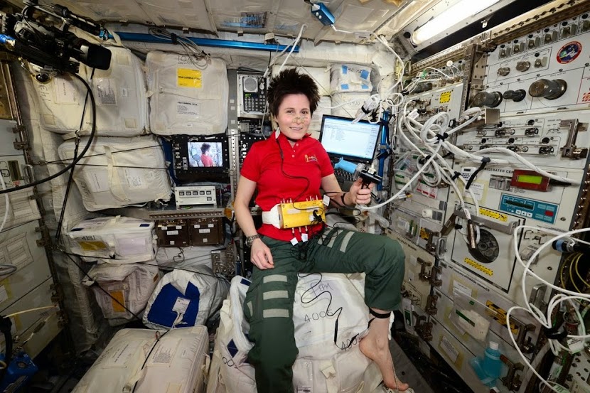
Samantha Cristoforetti űrhajós a Nemzetközi Űrállomáson dolgozik és a vénás keringést tanulmányozza - dr. Zamboni technológiáját használva - mindeközben a Földön sejtbiológusok az SM-es agyat vizsgálják. Az SM betegség érrendszeri kapcsolatának kutatása makro és mikrokozmosz szinten ezekben a percekben zajlik, a szemünk előtt!

Ezekben a percekben az űrben


Van videó is Samantháról amint a pletizmográfot használja és Zambonival kommunikál az űrállomásról!

Itt alant angolul olvasható Samantha beszámolója arról, hogyan használja Zamboni pletizmográfját az agyból a szív felé áramló vér mérésére. A képen a nyakára csatolva látható az a fekete gallér-szerű műszer amit remélhetőleg hamarosan magyar kutatók is a CCSVI mérésére, kutatására fognak használni.

Early in the morning on Monday it was also time for the first session of Drain Brain. Actually, we already had an ultrasound session early on in the mission, but for this particular set of measurements we had to wait for the replacement hardware to be delivered on Dragon, following the loss of the Orbital-3 mission. Specific instruments for Drain Brain include three strain-gauge pletismoghraphs, which look like collars of a stretchable material, as you can see in the picture.  They are actually sensors able to measure blood flow in the veins in a very simple and non-invasive manner, which is not dependent on the skills and interpretation of the operator, as is the case with ultrasound. While wearing these collars on my neck, arm and leg, I performed a series of breaths at 70% of my lung capacity, either remaining still or stretching and flexing my hand or my ankle. While doing that, I was breathing into our Pulmonary Function System and the software, via a graphic interface, was giving me instructions on when to start exhaling or inhaling. The main goal of the experiment is to study how the return of blood from the head to the heart changes in space, since we don’t have gravity effects helping with that. It’s something that we know little about for now and a better understanding of these circulatory mechanisms could potentially help in understanding some degenerative diseases of the brain. - Samantha Cristoforetti  forrás

Mindeközben a Földön:

Az új technológiák által az SM kutatók sejtszinten tudják vizsgálni az SM-es agyat, a gócok kialakulása ELŐTT. A következőkben három különösen fontos kutatást ismertetünk röviden.

A sejtbiológusok a legalacsonyabb szinten vizsgálják az SM-es agyat, sejtszinten. Ők nem azt vizsgálják, hogy bizonyos gyógyszerek hogyan hatnak, hanem azt kutatják hogy mi lehet az SM okozója. És mindegyikük lát kapcsolatot az SM, az érrendszer és az vér-agy gát sérülése között.

A NASA szeretné megérteni, hogy az űrhajósainak a 20%-a miért érkezik vissza a világűrből neurológiai és látásproblémákkal, és hogy ennek mi köze a vérkeringéshez. Ezért szakértőhöz fordultak.

http://www.nasa.gov/mission_pages/station/research/experiments/1278.html

Íme a vadonatúj kutatási eredmények, mind az SM és az érrendszer kapcsolatát erősítik. Az összefoglalókat angolul közlöm, mert sajnos nem látom értelmét magyarra fordítani - olyan kevesen érdeklődnek hazánkban eziránt a kutatási irány iránt. Szeretném hinni, hogy van akit érdekelnek az új kutatások és van aki még azt gondolja, hogy nem szabad kivárni azt a 20 évet, ami alatt ez a tudás mainstream tudássá változik, hanem kutatni kell és menni előre.

Elvégre is emberéletekről beszélünk.


1.  The Role of Angiogenesis in the Pathology of MS
http://www.ncbi.nlm.nih.gov/pmc/articles/PMC4253611/

Cell biologists from the University of Irvine have noted how the loss of endothelial tight junctions in the blood brain barrier contributes to inflammation and angiogenesis (the growth of new blood vessels) in the MS brain,  and how this process is initiated by hypoxia.  This low oxygen state and resultant angiogenesis occurs prior to formation of demyelinating lesions

This cellular research is further defining the hypothesis of cellular biologist Dr. Bernhard Juurlink, made in the 1990s.
http://ccsviinms.blogspot.com/2010/08/blood-flow-and-white-matter-lesions.html

It also fits in with the hypothesis of MS as a disease of hypoperfusion/reperfusion injury.
 http://ccsviinms.blogspot.com/2013/09/multiple-sclerosis-hypoperfusionreperfu.html


2. In vitro study of the direct effect of extracellular hemoglobin on myelin components.
http://www.ncbi.nlm.nih.gov/pubmed/25463632

The cellular biologists from the University of Guelph are looking at how blood particles damage myelin.  They are seeing microscopic deposits of hemoglobin in the MS brain, around the veins.  This blood contains iron, which when deposited into delicate brain tissue, begins a process of oxidative stress. 

"This study provides new insight into the mechanism by which hemoglobin exerts its pathological oxidative activity towards myelin components. This work supports further research into the vascular pathology in MS, to gain insight into the origin and role of iron deposits in disease pathogenesis, or in stimulation of different comorbidities such as cardiovascular disease."
This work confirms the theory of Dr. Zamboni from 2006, called his "Big Idea" theory, which saw the similarities of venous disease to MS, by noting how blood particles caused damage to tissue via the iron found in our red blood cells.   http://www.ncbi.nlm.nih.gov/pmc/articles/PMC1633548/
 
 
3.  Focal disturbances in the blood brain barrier are associated with formation of neuroinflammatory lesions
Neurobiologists from the University of Montreal are seeing changes to the blood brain barrier which happen before immune cells enter the MS brain. There are changes to the tight junctions of endothelial cells. 
 Our findings suggest that BBB breach occurs before significant immune cell infiltration and demyelination.
 
I wanted to briefly highlight these new studies, and encourage all readers to pursue cardiovascular and endothelial health in 2015.  
The discoveries of endothelial dysfunction and the link to the breakdown of the blood brain barrier in MS are being made.  While we wait for the venoplasty and pharmaceutical solutions, there is much that can be accomplished with lifestyle changes. 
http://ccsvi.org/index.php/helping-myself/endothelial-health


Joan Beal cikke alapján

Szólj hozzá!


nasazamboni.jpgFRISSÍTVE 2014 október 30

Sajnos a nemrég felrobbant NASA űrjármű vitte magával az űrbe Zamboni doktor felszerelését, mellyel az emberi vérkeringést szerették volna mérni az űrállomáson. Reméljük a CCSVI kutatására maradt még elég műszere. 

A NASA ugyanis Zamboni doktort bízta meg a vérkeringés kutatásával. Az általa a CCSVI kutatására fejlesztett nyaki pletizmográfot (régebben írtunk már róla) felviszik a Nemzetközi Űrállomásra, ahol a vérkeringést kutatják vele.

Talán sejt valamit az Öreg mégiscsak? :)


Szólj hozzá!

Miközben itthon teljes és dermedt csend van a CCSVI körül azóta, mióta ezt a blogot írom (2008 óta) Zamboni doktor nem pihen. Ez a bohókás olasz doktor képes volt a NASA után a Nature magazint is megbolondítani az elméleteivel, akik közölték a legújabb kutatását. CCSVI betegek vénáit vizsgálták meg egy új eszközzel (Synchrotron XRF analysis, egyfajta röntgen)  és kiderült, hogy sokkal több kalcium található ezekben a CCSVI betegek vénáiban, mint egy egészséges emberében. 

Hétköznapi nyelvre lefordítva: a felhalmozódott kalcium miatt a véna felépítése megváltozik, ami egyértelműen kimutatható a tanulmány szerint. A CCSVI egy létező jelenség, újabb tudományos tényekkel alátámasztva.

Izgalmas továbbá, hogy a tanulmány megemlít további neurológiai betegségeket, amik összefüggésben lehetnek a CCSVI-vel. Érdemes megnézni a cikket, nagyon látványos anyag.

1 komment

Frissítés 2014. október 4.

Kerri Cassidy az ausztrál CCSI szövetség elnöke egy ausztrál újságnak nyilatkozott az ausztrál kutatás eddigi eredményeiről:

A tervezett 160 betegből eddig 34-et vizsgáltak meg, akik közül 27-nél találtak CCSVI vénaszűkületet, ők részt is vettek a kettős vak tágítási procedúrán. Elég szignifikáns számok...

-----------------------

2012. július 24-én írtuk:

USALengyelországOlaszország, Bulgária, KuvaitSzlovákia, Szerbia és Csehország után Ausztráliában is megindultak a CCSVI műtétek, minden fentebb felsorolt országhoz hasonlóan egy kutatási program keretei között, állami támogatással és nem magán praxis keretei között.

Az ausztrál kutatás részletes paraméterei itt megtalálhatóak, a fontosabbakat összefoglalom:

  • 2 év alatt 160 beteget műtenek meg,
  • a kutatás randomizált, kettős vak és placebo kontrollált: a betegeket két csoportra osztják, minden résztvevőn angiogrammot (nyomásmérést) készítenek, de csak az egyik csoportot tágítják. 12 hónap múlva újra minden résztvevőn angiogramot végeznek és most a másik csoportot is tágítják, de sem az orvosok sem a beteg nem tudja, mikor tágították (a tágítás során valamilyen enyhe altatást alkalmaznak). 
  • A kutatást a The Alfred Hospital-ban végzik, Melbourne, Ausztrália.
  • A betegeket neurológusok vizsgálják és MSFC skálán mérik a változásokat.
  • A 160 önkéntes beteg pár nap alatt összegyűlt a kutatáshoz.

Ausztráliának kíváncsi, bátor és betegközpontú orvosai vannak, ezúton is teljes szívemből gratulálok nekik!

A kutatással kapcsolatos tudományos/szakmai kérdéseket a kutatás vezetőjének dr. Helen Kavnoudias -nak lehet címezni: 


Address: Radiology Research Unit, RAdiology Department
The Alfred
Level 1 Phillip Block, 55 Commercial Road
Melbourne, Vic, 3004
Country: Australia
Tel: +61 3 9076 3606
Fax: +61 3 9076 2153
Email: h.kavnoudias@alfred.org.au


Az Ausztrál CCSVI Facebook csoport elérhető itt. Szomorú és egyben felháborító olvasni hogy az ausztrál SM Kutatóközpont megígérte ugyan, de végül nem utalta el a kutatáshoz szükséges pénzt.


Címkék: kutatás ausztrália tanulmány sm vizsgálat orvosoknak

Szólj hozzá!

süti beállítások módosítása